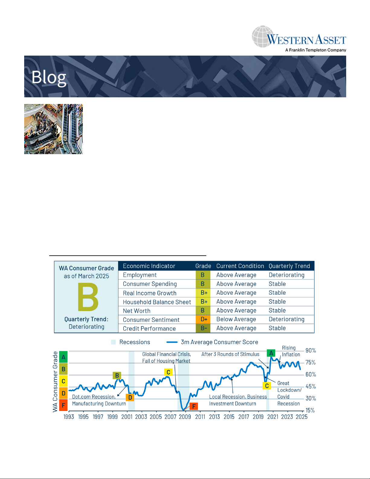
Wearable Sensors for Monitoring the Internal and External Workload of the Athlete PDF Free Download
1 / 19/19
100%

"*/63(83?644659(9,
&,9:,85!,9,8<,$50<,890:?
(9,&,9:,85!,9,8<,$50<,890:?(9,&,9:,85!,9,8<,$50<,890:?
"*/63(83?644659(9,&,9:,85"*/63(83?644659(9,&,9:,85
!,9,8<,$50<,890:?!,9,8<,$50<,890:?
(*;3:?"*/63(89/07
&,(8()3,",59689-68650:6805.:/,5:,85(3(5+>:,85(3&,(8()3,",59689-68650:6805.:/,5:,85(3(5+>:,85(3
&68236(+6-:/,:/3,:,&68236(+6-:/,:/3,:,
/8;<!",9/(+80
(9,&,9:,85!,9,8<,$50<,890:?
(4,9!!6=)6::64
(9,&,9:,85!,9,8<,$50<,890:?
188*(9,,+;
,3,9:,(80,3-,9
(9,&,9:,85!,9,8<,$50<,890:?
*49*(9,,+;
/809:0(5'684(5
(9,&,9:,85!,9,8<,$50<,890:?
*(@*(9,,+;
63058;4465+
(9,&,9:,85!,9,8<,$50<,890:?
*>+*(9,,+;
;:/689!+,5:0A,8
,3,9:,3-,9
/809:0(5'684(5
6336=:/09(5+(++0:065(3=6829(:/::79*644659*(9,,+;-(*;3:?=6829
0.0:(3
644659
,:=682
6.6
(8:6-:/,;8905.644659
!,*644,5+,+0:(:065!,*644,5+,+0:(:065
",9/(+80!0!#%669,:(3&,(8()3,9,59689-684650:6805.:/,05:,85(3(5+,>:,85(3=68236(+
6-:/,(:/3,:,5710.0:,+/::79+6068.9
#/098:0*3,09)86;./::6?6;-68-8,,(5+67,5(**,99)?"*/63(83?644659(9,&,9:,85!,9,8<,$50<,890:?
:/(9),,5(**,7:,+-6805*3;906505(*;3:?"*/63(89/07)?(5(;:/680@,+(+40509:8(:686-"*/63(83?644659
(9,&,9:,85!,9,8<,$50<,890:?68468,05-684(:06573,(9,*65:(*:+0.0:(3*644659*(9,,+;
&!$(;:/689/(<,4(+,:/09=682-8,,3?(<(03()3, 3,(9,:,33;9/6=:/09(**,99/(9),5,A:,+68047(*:,+?6;

REVIEW ARTICLE OPEN
Wearable sensors for monitoring the internal and external
workload of the athlete
Dhruv R. Seshadri
1
, Ryan T. Li
2
, James E. Voos
3
, James R. Rowbottom
4
, Celeste M. Alfes
5
, Christian A. Zorman
6
and
Colin K. Drummond
1
The convergence of semiconductor technology, physiology, and predictive health analytics from wearable devices has advanced its
clinical and translational utility for sports. The detection and subsequent application of metrics pertinent to and indicative of the
physical performance, physiological status, biochemical composition, and mental alertness of the athlete has been shown to reduce
the risk of injuries and improve performance and has enabled the development of athlete-centered protocols and treatment plans
by team physicians and trainers. Our discussions in this review include commercially available devices, as well as those described in
scientific literature to provide an understanding of wearable sensors for sports medicine. The primary objective of this paper is to
provide a comprehensive review of the applications of wearable technology for assessing the biomechanical and physiological
parameters of the athlete. A secondary objective of this paper is to identify collaborative research opportunities among academic
research groups, sports medicine health clinics, and sports team performance programs to further the utility of this technology to
assist in the return-to-play for athletes across various sporting domains. A companion paper discusses the use of wearables to
monitor the biochemical profile and mental acuity of the athlete.
npj Digital Medicine (2019) 2:71 ; https://doi.org/10.1038/s41746-019-0149-2
INTRODUCTION
Technological advancements have enabled athletes, coaches, and
physicians to track functional movements, workload, biomecha-
nical and bio-vital markers utilizing wearable sensors to maximize
performance and minimize the potential for injury.
1–3
Wearable
monitoring systems can provide continuous physiological data thus
permitting the development of accurate treatment plans and
player-specific training programs to potentially mitigate and
alleviate injuries.
4
Herein, we define a wearable device as a sensor
or sensor suite unencumbered by wires for the continuous and
non-invasive detection of biosignals, analytes, or biomechanical and
impact forces for monitoring human health and performance. Over
the past two decades, the wearables field has moved from a device
to a systems viewpoint, where the system combines the device with
analytics. While previous literature has reviewed specifictechnical
domains of the wearables field, such as sensors,
5–8
materials,
9–12
and soft interfaces
13–15
or focused on the fabrication and
application of such devices to address a specific medical condition,
such as atrial fibrillation,
16–18
cystic fibrosis,
19–21
or diabetes,
22–27
there remains an unmet medical need to assess, develop, and
validate this technology specifically for sports medicine. Given the
heightened attention to athlete safety and performance, this review
evaluates the translational utility of wearable devices to detect key
metrics pertinent to human performance assessment (Fig. 1).
The organization of this review is structured around discussing the
value wearable sensors provide in sports to monitor player activity
levels and mitigate injury. We progress through this review by
discussing the utility of wearable sensors in two domains crucial to
human performance ranging from an athlete’s physical performance
and physiological status. Our goal in each of these areas is centered
around reviewing both scientific literature and current commercially
available devices to provide a comprehensive view of wearables for
sports medicine (Tables 1–5). This review is specifically targeted
towards those whose interests lie in the application and translation of
wearable sensors for assessing human performance.
PHYSICAL PERFORMANCE AND SAFETY OF THE ATHLETE
Position and motion
The ability to monitor position and movement profiles of an
athlete is critical in developing improved training regimens to
maximize individual performance (Fig. 2). The accuracy of devices,
such as pedometers has been in question and was recently
studied. Researchers compared the accuracy of the “step-count”
feature between dedicated smartphone-based pedometer appli-
cations (Galaxy S4 Moves App, iPhone 5s Moves App, iPhone 5s
Health Mate App, iPhone 5s Fitbit App) and wearable devices
(Nike Fuelband, Jawbone UP24, Fitbit Flex, Fitbit One, Fitbit Zip,
and Digi-Walker SW-200) with direct observation of step counts.
28
Results showed a relative difference between actual and reported
mean step count of −0.3% to 1.0% for pedometers and
accelerometers, −22.7% to −1.5% for wearable devices, and
−6.7% to 6.2% for smartphone applications. Such differences were
attributed to the robustness of the IC technology and software
algorithms used to determine a step. Step counts are often used
to derive other measures of physical activity, such as distance
Received: 19 January 2018 Accepted: 8 July 2019
1
Department of Biomedical Engineering, Case Western Reserve University, 10900 Euclid Avenue, Cleveland, OH 44106, USA;
2
Department of Orthopaedic Surgery, University
Hospitals Cleveland Medical Center, Cleveland, OH 44106, USA;
3
University Hospitals Sports Medicine Institute, Cleveland, OH 44106, USA;
4
Department of Cardiothoracic
Anesthesiology, The Cleveland Clinic, 9500 Euclid Avenue, Cleveland, OH 44195, USA;
5
Frances Payne Bolton School of Nursing, Case Western Reserve University, 9501 Euclid Avenue,
Cleveland, OH 44106, USA and
6
Department of Electrical Engineering and Computer Science, Case Western Reserve University, 10900 Euclid Avenue, Cleveland, OH 44106, USA
Correspondence: Dhruv R. Seshadri (Dhruv.Seshadri@case.edu)
www.nature.com/npjdigitalmed
Scripps Research Translational Institute

traveled or calories expended.
28
Hence, improving measurement
accuracy is crucial to measure and appropriately tailor workout
regiments for elite-level athletes.
Movement-based sensors currently in use for sports-medicine
include accelerometers and global positioning satellite (GPS)
devices, often used in combination (Table 1). Accelerometers
generate highly accurate analyses of movement with high sampling
rates and have been included in wrist-based devices, such as the
Nike Fuelband, Jawbone UP, and Microsoft Band. This technology
has been widely adopted in the sporting community ranging from
Australian Football,
29
Rugby,
30,31
NFL,
32
National Hockey League
(NHL),
33
and swimming.
34–36
Energy expenditure can be determined
from tri-axial accelerometers via the integration of acceleration over
time.
37,38
The determination of energy expenditure, position,
movement, and balance control during practices or games has
shown to be instrumental in tailoring the training regimen of
athletes to minimize the incidence of soft tissue injuries.
Banister et al. postulated that athletic performance can be
estimated as a function of fatigue and fitness
39
(Fig. 2). Building
upon this model, Morton et al. suggested that an opportune
training stimulus is one that maximizes performance by utilizing
an appropriate training load, while simultaneously minimizing
injury and fatigue.
40
A working definition of fatigue is “any
exercise-induced or non-exercise-induced loss in total perfor-
mance due to various physiological factors, athlete reported
psychological factors, or a combination of the two”.
41
It is well
known that fatigue decreases athletic performance and that
training induces numerous neurophysiological and psychological
changes in an athlete’s body. There are two forms of fatigue:
central fatigue and peripheral fatigue. Central fatigue is the fatigue
resulting from the central nervous system (CNS) and the
transmission of signals from the brain to the muscle.
42
Central
fatigue is related to the interaction between the brain and the
spinal cord.
43
Researchers have hypothesized that the differentia-
tion between a good athlete and an elite-level athlete is their
individual ability to ignore such neurotransmissions during high-
acuity situations (e.g. high profile matches or workouts).
42
Peripheral fatigue is the failure to maintain an expected power
output caused by the depletion of glycogen, phosphate
compounds, or acetylcholine within the muscular unit or by the
accumulation of lactate or other metabolites that are released
during activity.
44,45
Peripheral fatigue occurs within the muscle
and can be thought of as ‘muscle fatigue’.
43
As such, wearable
sensors can be used to measure parameters indicative of the
peripheral fatigue of the athlete, as is discussed in detail
throughout this review. For simplicity purposes, we refer to
peripheral fatigue as simply fatigue.
Monitoring internal (e.g. physiological or perceptual ‘response’)
and external training loads (e.g. physical ‘work’) can enable sports
trainers and clinicians to assess the fatigue and fitness levels of
athletes in real time. Internal workload includes the session rate of
perceived exertion (sRPE) and heart rate.
46
At the completion of
each training session, athletes provide a 1–10 ‘rating’based on the
intensity of the session.
46
The intensity of the session is multiplied
by the session duration to provide the internal training load.
46
The
product can be thought of as the athletes’“exertional minutes”.
46
Advancements in MEMS fabrication techniques and device
packaging have allowed for the detection of multi-axial move-
ment to calculate an external training load (e.g. PlayerLoad™
3
).
External workload can be thought of as how much load is placed
on the body and can be quantified using torso-mounted wearable
devices which contain a GPS and a tri-axial accelerometer.
46
PlayerLoad™can be calculated via the instantaneous rate of
change of acceleration. Accumulated PlayerLoad™can be
calculated as the summation of PlayerLoad™over the desired
time interval (usually over a span of 1–7 days).
Metrics such as total distance run, weight lifted, number and
intensity of sprints or collisions can be determined using GPS-
based sensors. Position sensors triangulate signal transmission
from multiple GPS satellites orbiting the earth and can accurately
determine the velocity and position (within 1 m) of an athlete on a
field. These devices are playing an instrumental role in sports
performance analysis by allowing coaches, physicians, and trainers
to better understand real-time physical demands of an ath-
lete.
30,37,47
GPS silicon chips combined with tri-axial acceler-
ometers have been used to record physical activities during
different times of the day and for specific position groups on a
team.
48
The majority of work to assess human motion and its
correlation to sports performance has involved the use of
commercial GPS-based devices, such as the Catapult devices
(OptimEye S5) and Zebra Technologies GPS device. The Catapult
product, for example has a fully packaged processing IC,
accelerometer, gyroscope, and magnetometer to measure body
position, impact forces, velocity, acceleration, and direction in a
continuous manner.
49
In a study utilizing the Catapult OptimEye
S5 and video tracking technology, 20 professional Australian
Football League (AFL) players were studied during four in-season
matches to describe and quantify the frequency, velocity, and
acceleration at impact during tackling
29
(Fig. 3a–c). Distributions in
tackles were quantified and classified as a function of percent
distribution of tackles versus player load (Fig. 3a), player velocity
versus tackle intensity (Fig. 3b), and player load versus tackle
intensity (Fig. 3c). Differences in accelerometer data between
tackles were observed to be progressively greater in intensity
thereby providing support for the use of accelerometers to assess
impact forces in contact-based sports.
29
In another study, GPS
sensors and related analytics were used by National Collegiate
Athletic Association (NCAA) Division I Football athletes to record
workload, velocity, distance, and acceleration during both
practices and games.
48,50
The studies found significant variation
in movement profiles among collegiate football players and the
authors identified the need for position-specific and game-specific
physical conditioning strategies to maximize player performance,
limit the effects of fatigue, and minimize the onset of injuries.
Fig. 1 Four areas of focus as it relates to assessing human
performance. The central theme of this review is the use of
wearable sensors to maximize the performance and safety of the
athlete. This involves the detection and measurement of the internal
and external workload of the athlete which are based on the
athlete’s physical performance, physiological status, biochemical
composition, and mental acuity
D.R. Seshadri et al.
2
npj Digital Medicine (2019) 71 Scripps Research Translational Institute
1234567890():,;

Table 1. Examples of wearable technology companies with products applicable towards assessing the position and motion of the athlete
Company Sampling of products Product type Product functionality Headquarters
Adidas miCoach Fit Smart, miCoach
Smart Run
Watch Heart rate, GPS, distance Herzogenaurach, Germany
Apple Apple Watch Watch Heart rate, distance, email, ECG, text messages, phone Cupertino, CA
BioSensive Technologies Inc. Joule Earrings Heart rate, calories burned, steps taken, overall activity level Ontario, Canada
Catapult OptimEye S5, Vector Device unit Movement, Turn rates, orientation, heart rate. Device placed below
the neck (tucked in shoulder pads)
Melbourne, Australia
Fitbit Flex, One, Alta Watch Steps walked, distance, heart rate, sleep quality, pedometer,
calories burned
San Francisco, CA
Garmin Vivoactive, Vivosmart, Vivofit Watch Pedometer, sleep quality, heart rate, distance Schaffhausen, Switzerland
Jabra Sports Pulse Wireless
Headphone
Headphone Accelerometer and heart rate monitoring Ballerup, Denmark
Jawbone Up Band Pedometer, distance, heart rate, sleep quality, calories San Francisco, CA
Karacus Polaris, Zeta, Proxima Watch Movement, phone, email Chapel Hill, NC
Kitman Labs Capture Sensor mounted on
computer
Biometric data via machine learning, GPS, and player tracking Dublin, Ireland
Microsoft Microsoft Band Band Heart rate, calories burned, sleep quality, email, text Redmond, WA
Nike Fuelband Band Pedometer, GPS Washington County, Oregon
Polar A360, Loop Crystal, Loop 2 Band Heart rate, performance tracker Kempele, Finland
Samsung GearFit 2 Watch GPS, sleep, heart rate, calories, pedometer Seoul, South Korea
Sansible Technologies LiveSkin Textile electronics Speed, impact, position Edinburgh, United Kingdom
Starkey Hearing Technologies Livio AI Hearing Aid Hearing aid Translates foreign languages, contains a pedometer, tracks physical
activity (wellness score)
Eden Prairie, MN
Stifit Stifit band Band Blood oxygen, body mass index, calories burned, distance, fatigue,
heart rate, sleep
San Francisco, CA
TruSox TruSox Socks Non-slip socks to generate greater speed and agility Baltimore, MD
Under Armor HTC Grip Wristband Heart rate, calories burned, distance traveled Baltimore, MD
Vert G-Vert, VERT Coach Device unit Measure G-force in movement, acceleration, kinetic energy, power Fort Lauderdale, FL
Vibrado Technologies Vibrado Technologies Textile electronics Accelerometer to measure shot angle, arm height, release point.
Sleeve to be worn over forearm
Sunnyvale, CA
Zebra Technologies Zebra Tracking Device Device unit RFID used to quantify movement and distance profiles. Device
placed below the neck in shoulder pads or sewn into jersey
Lincolnshire, IL
Zephyr BioHarness 3, HxM™Smart,
HxM™BT
Textile Electronics Heart rate, respiration, tri-axial accelerometer, heart rate, activity,
posture, oxygen saturation levels
Annapolis, Maryland (Founded in
New Zealand)
Data for this table was acquired from company websites and social media sites affiliated with each company
D.R. Seshadri et al.
3
Scripps Research Translational Institute npj Digital Medicine (2019) 71

The combination of the internal and external workloads of the
athlete determine the training outcome.
46
An athlete’s internal or
external workload can be computed over a 1-week period (acute
workload) and over a 3–4-week period (chronic workload). Work
by Gabbett suggested that the ratio of the acute-to-chronic
workload, herein referred to as “ACWR”, can be used to determine
if an athlete is overtraining, undertraining, or training at the
opportune intensity
46
(Fig. 2b). Furthermore, Gabbett showed that
calculation of this ratio enables sports scientists to predict the
chance an athlete suffers an injury as a result of improper load
management.
46
Deriving this ratio provides an index of athlete
preparedness and considers the training load that the athlete has
performed relative to the training load that the athlete has been
prepared for.
51
The use of the ACWR emphasizes both the positive
and negative consequences of training. The first study to
investigate the relationship between ACWR and the risk of injury
was performed on elite cricket fast bowlers.
52
Training loads were
estimated from both sRPE and balls bowled. When the acute
workload was similar to or lower than that of the chronic workload
(e.g. ACWR ≤0.99), the likelihood of injury for fast bowlers in the
next 7 days was 4%.
52
However, when the ACWR was ≥1.5 (e.g.
workload in the current week was 1.5 times greater than what the
bowler was prepared for), the risk of injury was 2–4 times greater
in the subsequent 7 days.
52
While such observations are indicative
of the sport being studied, until more robust data sets are
available, caution must be heeded when applying these
recommendations to individual sport athletes. Despite this, a
general trend can be concluded. If the acute training load is low
(e.g. the athlete is experiencing minimal fatigue) and the rolling
average (RA) chronic training load is high (e.g. the athlete has
developed ‘fitness’), then the athlete will be in a well-prepared
state and thus, the ACWR will be ≤1.
46
If the acute load is high (e.g.
training loads have been rapidly increased resulting in ‘fatigue’)
and the RA chronic training load is low (e.g. the athlete has
performed inadequate training to develop ‘fitness’), then the
athlete will be in a fatigued state and thus, the ACWR will be ≥1.
46
In terms of injury risk, ACWRs within the range of 0.8–1.3 could be
considered the training ‘sweet spot’, while an ACWR ≥1.5 could
represent the ‘danger zone’.
46
The RA model
53
(Eqs. (1–4)) and exponentially weighted moving
average (EWMA) model
54
(Eqs. (5–10)) are two methods used to
calculate the training load of the athlete with or without the use of
wearable sensors like the Catapult OptimEye S5 (Eqs. (11–14)).
3
The RA model uses an absolute (i.e. total) workload performed in
one week (acute workload) relative to the 4-week chronic
workload (e.g. 4-week average acute workload).
53
Equation (1)
Table 2. Examples of wearable technology companies for impact monitoring
Company Sampling of products Product type Product functionality Headquarters
2ND Skull Cap, Band Garment Polyurethane-based composite dissipates impact Pittsburgh, PA
Athlete Intelligence Vector Mouthguard,
Shockbox®sensor
Mouth guard Tracks linear and rotational accelerations of head
impacts
Kirkland, WA
BrainScope Ahead 300 Hand-held point of
care device
Disposable electrode sensors to detect head
injuries
Bethesda, MD
Force Impact
Technologies
Fitguard™Mouth guard Embedded sensors relate collision intensity via
color coded LED’s on the front of the
mouth guard
Los Angeles, CA
Tempe, AZ
Hiji Hiji Band Head band Impact forces, intensity Phoenix, AZ
Jolt Jolt Sensor Sensor Impact forces, Concussion monitoring. Sensor
clipped to garment
Boston, MA
Mamori Mamori Mouth guard Inertial sensors measure impact forces on
the head
Dublin, Ireland
Noggin Pro Noggin, Noggin Pro Skull caps Gel capsules in skull cap dissipate forces
from skull
Toronto, ON
Performance
Sports Group
Q-Collar Neck collar Concussion prevention by applying pressure on
the jugular vein
Cincinnati, OH
X2 Biosystems X-Patch Pro Flexible sensor Tri-axial accelerometers to measure impact Seattle, WA
X2 Mouthguard
Data for this table was acquired from company websites and social media sites affiliated with each company
Table 3. Examples of wearable technology companies for monitoring the biomechanical forces on the athlete
Company Sampling of products Product type Product functionality Headquarters
CricFlex CricFlex Sleeve Measures arm angle and force during bowling Islamabad, Pakistan
Heddoko Heddoko Smart
Garment
Biomechanics of movement, deviation from
benchmarks and movement standards, injury risk
Montreal, Canada
Motus Global mThrow™, motusPro™Sleeve Accelerometer to measure joint angles, velocity,
stress, strain
Massapequa, New York
Ft. Lauderdale, FL
Protonics Technologies Protonics T2 Device Offsets left-right biomechanical imbalance to reduce
muscle pain. Attached to left leg
Lincoln, NE
Data for this table was acquired from company websites and social media sites affiliated with each company
D.R. Seshadri et al.
4
npj Digital Medicine (2019) 71 Scripps Research Translational Institute

Table 4. Examples of wearable technology companies with products applicable towards monitoring heart rate and muscle oxygen saturation
Company Sampling of products Product type Product functionality Headquarters
1st Round Athletics EnergyDNA™Body suit Converts heat to IR which expands blood vessels for greater blood flow Los Angeles, CA
Athos Athos Wearables Vest and pant Muscle activity and heart signals via EMG San Francisco, CA
Hexo Skin Astroskin, Smart Kit Sleeve Cardiac frequency, respiratory rate and volume, sleep, acceleration Montreal, Canada
San Francisco, CA
Huawei Honor Band A1 Watch Cardio-respiratory fitness Shenzen, China
Humon Hex Device unit Non-invasive measurement of O
2
content in muscles Boston, MA
Komodo Technologies Inc. AIO Smart Sleeve Sleeve ECG, heart rate, sleep analysis Winnipeg, Canada
Kymira Kymira Sports T-Shirt Smart garments for cardiac monitoring to prevent heart attacks in athletes Reading, United Kingdom
LifeBeam LifeBeam baseball cap SmartHat Embedded sensors measure heart rate and calories New York, NY
MC10 BioStamp RC™, BioStamp nPoint®,
Kintinuum
Epidermal sensor BioStamp RC™: activity, cardiac activity, EMG, and posture Boston, MA
BioStamp nPoint®: activity, posture EMG, and sleep metric, vital signs
Kintinuum: quantify treatment efficacy
Myovolt Myovolt Sleeve EMG to increase circulation, boost muscle power Hong Kong, China
Rotex Technologies roMage, roSport, roCare, roFashion Electronic tattoo roMage: brain and muscle control, gesture recognition Austin, TX
roSport: blood O
2
saturation, heart rate, skin hydration
roCare: blood pressure, ECG, respiration, skin hydration, temperature
roFashion: glows on skin (fashion purposes)
Spire Spire Device unit Measures respiration to quantify and detect stress, calorie tracking,
pedometer
San Francisco, CA
Withings Steel HR, Activité, Go, Pulse O
2
Bands Heart rate, distances, email, text messages, phone Issy-les-Moulineaux, France
Xiaomi Mi Band Band Time, pedometer, heart rate Beijing, China
Xmetrics Xmetrics Pro, Xmetrics Fit Device unit Calories, strokes taken, laps swam, pace Milan, Italy
Data for this table was acquired from company websites and social media sites affiliated with each company
D.R. Seshadri et al.
5
Scripps Research Translational Institute npj Digital Medicine (2019) 71

represents the exertional minutes per workout which is the
product of the session rating of perceived exertion and the
duration of the workout in minutes. The sRPE is a scale from 1 to
10 with progressing intensity of the workout deemed by the
athlete and training staff. Equation (2) shows the acute player load
(PL) which is the summation of the exertional minutes per
workout for a given week (e.g. from day 1 to day 7). For the sake of
simplicity, we assume the athlete is completing one workout
per day. Equation (3) shows the chronic PL which is computed by
taking the average of the acute PL over the duration of weeks
(denoted as n). Equation (4) shows the ACWR which is the ratio
between the acute PL for the given week (Eq. (2)) and the chronic
PL (calculated from Eq. (3)). The RA model suggests that each
workload in an acute and chronic period is equal. In other words,
the model considers there to be a linear relationship between load
and injury. The assumption in this model is that all workload in a
given time period is equivalent. Key drawbacks of this model are
that the model does not account for any decays in fitness and it
does not accurately represent variations in the manner in which
loads are accumulated.
Exertional minutes per workout ¼SRPEðÞ´duration of workout in minutesðÞ
(1)
Acute PL ¼XD¼7
D¼1exertional minutes per workout (2)
Chronic PL ¼PW¼n
W¼1Acute PL
n(3)
ACWR ¼Acute PL for given week
Chronic PL (4)
Table 5. Examples of wearable technology companies for monitoring sleep
Company Sampling of products Product type Product functionality Headquarters
EmfitEmfit QS Device unit Tracks sleep by monitoring movement and heart rate Vaajakoski, Finland
Kokoon Kokoon EEG Headphones Movement and EEG sensors determine relaxation and sleep quality Limerick, Ireland
Moov Moov Wrist-based device Heart rate, sleep quality, and activity tracker San Francisco, CA
WHOOP WHOOP Band Wrist-based device Heart rate, body temperature, movement, and sleep Boston, MA
Data for this table was acquired from company websites and social media sites affiliated with each company
Fig. 2 Value proposition of wearable sensor technology to monitor athlete training load to minimize soft-tissue injuries. aHypothetical
relationship between training loads, fitness, injuries, and performance. Inadequate and excessive training loads could result in increased
injuries, reduced fitness, and poor team performance. bInterpreting and applying ACWR data to predict the likelihood of subsequent injury.
The green-shaded area (‘sweet spot’) represents the ACWR where the risk of injury is low. The red-shaded area (‘danger zone’) represents the
ACWR where the risk of injury is high. To minimize the risk of injury, athletes should aim to maintain their ACWR within a range of ~0.8–1.3.
cAthlete workout can be monitored via workout logs and self-tracking methods, assessing the sRPE levels, or using wearable technology to
quantify movement parameters. The application of wearable sensors to monitor athletic performance and training has provided an added
advantage compared to current and past methods by enabling sports scientists and clinicians to quantify the workout, to calculate the ACWR,
and to predict the onset of injury. Figure was adapted and modified from Gabbett et al.
46
a,b
D.R. Seshadri et al.
6
npj Digital Medicine (2019) 71 Scripps Research Translational Institute

Sports scientists have started to apply the EWMA model to
circumvent the drawbacks posed by the RA model.
54
The EWMA
model places a greater weight on the most recent workload an
athlete has performed by assigning a decreasing weighting for
each older workload value (time decay constant, λ
a
) and the non-
linear nature of injury occurrence and workload.
54
Equation (5)
shows the exertional minutes per workout which is the product of
the session rating of perceived exertion and the duration of the
workout in minutes. The sRPE is a scale from 1 to 10 with
progressing intensity of the workout deemed by the athlete and
training staff. Equation (6) shows the degree of decay, λ
a
, which is
a value between 0 and 1, with higher values of λ
a
discounting
older observations in the model at a faster rate. In the following
equation, nrepresents the time decay constant. Equation (7)
shows the formula to calculate the EWMA for a given day which is
based on the exertional minutes, calculated from Eq. (5), the
degree of decay from Eq. (6), and the EWMA from the preceding
day. Equation (8) shows the acute player load (PL) which is the
summation of the EWMA for a given week (e.g. from day 1 to day
7). For the sake of simplicity, we assume the athlete is completing
1 workout per day. Equation (9) shows the chronic PL which is
computed by taking the average of the acute PL over the duration
of weeks (denoted as n). Equation (10) shows the ACWR which is
the ratio between the acute PL for the given week (Eq. (8)) and the
chronic PL (calculated from Eq. (9)). A recent study sought to
investigate if any differences existed between the RA and EWMA
models pertaining to ACWR calculation and subsequent injury risk
in elite Australian footballers.
54
Fifty-nine athletes from an AFL
club participated in this 2-year study. A total of 92 individual
sessions were recorded. Each season consisted of a 16-week
preseason phase comprised of both running and football-based
sessions, followed by a subsequent 23-week in-season competi-
tive phase. The Catapult OptimEye S5 GPS sensor, sampled at
10 Hz, was used to quantify training and match workloads of
players. The triaxial accelerometer, gyroscope, and magnetometer
were each sampled at 100 Hz. The study demonstrated that a high
ACWR was significantly associated with an increase in injury risk
for both models. The EWMA model had significantly greater
sensitivity to detect increases in injury likelihood at higher ACWR
ranges during both the preseason and in-season periods. The
study concluded that the EWMA model may be better suited to
modeling workloads and injury risk than the RAs than the ACWR
Fig. 3 Wearable sensors monitor the biomechanical performance of the athlete. aDistribution of tackles (n=352) made and against peak
instantaneous Player Load™.bPeak velocity for tackles made and against associated with tackle intensity categorized as low (n=115),
medium (n=216), and high (n=21). cPeak Player Load™for tackles made and against associated with tackle intensity categorized as low (n
=115), medium (n=216), and high (n=21). dRelative displacements of the mouthguard sensor from the skull studied using high speed
video. Among 16 trials, the mouthguard always had the smallest (sub-millimeter) displacement from the skull, within video error, compared to
the skull cap and skin patch. eRelative displacements of the Reebok skull cap from the skull studied using high speed video. fRelative
displacements of the xPatch Gen2 skin patch sensor from the skull studied using high speed video. gmotusBASEBALL sensor exhibited higher
peak elbow valgus torque for baseball pitching compared to football throwing. Data demonstrates the utility of the sensor to measure
biomechanical forces during non-stationary periods on an athlete. hmotusBASEBALL sensor used to quantify the average valgus torque on
the elbow for baseball pitching and football throwing between foot contact and maximum internal rotation. “
a
Significantly different (p< 0.01)
from Low;
b
significantly different (p< 0.01) from Medium. No significant differences between tackles made and against.”Figures were
reproduced with permission from Gastin et al.
29
a–c, Wu et al.
79
d–f, and Laughlin et al.
89
g–h
D.R. Seshadri et al.
7
Scripps Research Translational Institute npj Digital Medicine (2019) 71

model. Regardless of the ACWR model utilized, spikes in acute
workload were significantly associated with an increase in injury
risk.
Exertional minutes per workout ¼SRPEðÞ´duration of workout in minutesðÞ
(5)
λa¼2
Nþ1;where 0<λa<1(6)
EWMAtoday ¼Exertional minutes per workoutðÞλa
ðÞþ 1λa
ðÞEWMAyesterday
(7)
Acute PL ¼XD¼7
D¼1EWMAtoday (8)
Chronic PL ¼PW¼n
W¼1Acute PL
n(9)
ACWR ¼Acute PL for given week
Chronic PL (10)
Wearable sensors are currently being used to minimize injury in
professional football via careful monitoring of training load and
other biometrics during the rehabilitation period (Fig. 2c). The
variability of GPS data and accelerations of the torso have been in
question when it comes to monitoring the loads of the lower
limbs. This is because distance traveled and velocity do not
represent the mechanical load experienced by the musculoske-
letal tissue. This is specifically relevant in sports such as basketball,
which are constrained to a small-space, where players experience
high loads of physical stress by performing explosive jumping and
landing activities, which are not accurately captured by distance,
speed, or torso athlete movement analysis systems.
55,56
To
mitigate such issues, the Zebra GPS device and Catapult OptimEye
S5, both of which are considered the most accurate wearable
devices in sports today, are housed in player tracking devices in an
attempt to negate some of the aforementioned issues. Addition-
ally, the Catapult device has shown to mitigate such issues by
incorporating tri-axial movements into their analytical models to
accurately calculate PlayerLoad™from their sensor.
3
The Zebra
GPS device is currently approved by the NFL for use to track player
movement and has been utilized by select teams to monitor
training loads.
57
Equation (11) provides the analytical platform of
the Catapult OptimEye S5 which utilizes a tri-axial accelerometer
to calculate PlayerLoad™(PL) based on acceleration in the x,y, and
zdirections. Equation (12) shows the summation of PL from the
initial to end time of interest (in most cases this is from the start to
the end of a training session) denoted as AccPL™. Equation (13)
shows how the RA model can be used to calculate Acute PL,
analogous to Eq. (2). However, in this case, PL is calculated from
Eq. (11) using a wearable sensor. Eq. (14) shows how the EWMA
model can be used to calculate PL for the given day using PL
calculated from a wearable sensor. The ACWR can be calculated
utilizing either model, adapting the set of equations presented
(rolling average, Eqs. (1–4); EWMA, Eqs. (5–10).
PL ¼ffiffiffiffiffiffiffiffiffiffiffiffiffiffiffiffiffiffiffiffiffiffiffiffiffiffiffiffiffiffiffiffiffiffiffiffiffiffiffiffiffiffiffiffiffiffiffiffiffiffiffiffiffiffiffiffiffiffiffiffiffiffiffiffiffiffiffiffiffiffiffiffiffiffiffiffiffiffiffiffiffiffiffiffiffiffiffiffiffiffiffiffiffiffiffiffiffiffiffiffiffiffiffiffiffiffiffiffiffiffiffiffiffiffiffiffiffiffiffiffiffiffiffiffiffiffiffi
fwdt¼iþ1fwdt¼i
ðÞ
2þsidet¼iþ1sidet¼i
ðÞ
2þupt¼iþ1upt¼i
2
q
(11)
AccPL ¼Xt¼n
t¼0ffiffiffiffiffiffiffiffiffiffiffiffiffiffiffiffiffiffiffiffiffiffiffiffiffiffiffiffiffiffiffiffiffiffiffiffiffiffiffiffiffiffiffiffiffiffiffiffiffiffiffiffiffiffiffiffiffiffiffiffiffiffiffiffiffiffiffiffiffiffiffiffiffiffiffiffiffiffiffiffiffiffiffiffiffiffiffiffiffiffiffiffiffiffiffiffiffiffiffiffiffiffiffiffiffiffiffiffiffiffiffiffiffiffiffiffiffiffiffiffiffiffiffiffiffiffiffi
fwdt¼iþ1fwdt¼i
ðÞ
2þsidet¼iþ1sidet¼i
ðÞ
2þupt¼iþ1upt¼i
2
q
(12)
Acute PL ¼XD¼7
D¼1AccPL (13)
EWMAtoday ¼PLðÞλa
ðÞþ 1λa
ðÞEWMAyesterday
(14)
In a specific example reported by an American sporting
network, the device was used to accurately track the recovery of
an athlete after the individual suffered a season ending injury the
previous year.
57
The sensor was placed underneath the shoulder
pads (analogous to that of the Catapult device) or sewn into the
jersey to generate biometric measurements, such as movement
profiles and workload to gauge the athlete’s performance and
workload during recovery relative to his peak performance and
workload prior to the injury. Additionally, utilizing the Catapult
OptimEye S5 wearable sensor, authors of this review have recently
studied the effects of player workload on soft tissue injuries in a
single NFL team over two seasons.
32
Rapid changes in workload
over a one-week period when compared to the average workload
over a month were associated with a significant increase in risk of
hamstring and other soft tissue injuries. The studies demonstrated
that monitoring athletic training programs during the pre-season
compared to the post-season utilizing wearable technology have
assisted team athletic trainers and medical staff in developing
programs to optimize player performance and minimize soft-
tissue injuries.
32
Impact detection
The spongy nature of a woodpecker’s skull acts like a shock
absorber by pinching the jugular vein to increase blood pressure
in the brain to protect it from the 12,000 daily hammerings it
performs on trees.
58
Unfortunately, humans do not have any sort
of ‘protection mechanism’to mitigate or dissipate impact forces
on the brain.
58
The onset of concussions, brain injuries, and
mental health illnesses caused by repeated trauma to the head
have paved the way for newer technologies to detect and
eliminate chronic traumatic encephalopathy (CTE).
59
CTE is a
neurodegenerative disease found in individuals who have
experienced repeated traumatic brain injury (TBI) or concussions.
In these conditions, stretching, compression, and shearing of
axons during sudden brain movements over extended periods are
hypothesized to cause axonal injury.
60
The high incidence of such
injuries in athletes is of major concern in modern collision sports.
61
The American Academy of Neurology (AAN) defines a concussion
as a “pathophysiologic disturbance in neurologic function
characterized by clinical symptoms induced by biomechanical
forces”.
62
Guskiewicz et al. concluded that former NFL and
collegiate football players who reported multiple concussions
were at higher risk for depression and memory loss.
63,64
Research
on concussions and CTE is still rudimentary and primarily
supported by clinical models.
59
There remains a strong clinical
need to develop devices, which could quantitatively and
qualitatively measure impact forces on the brain to decrease the
onset of concussions and reduce the incidence of CTE. Currently,
work is being done to design custom personal protection
equipment (PPE), such as helmets and mouthguards to improve
player safety.
65
Research by Stenger et al. and McCrory et al.
showed the potential applicability of mouth guards towards
preventing head and spinal injuries.
66,67
Companies such as i1
Biometrics, Mamori, and Force Impact Technologies have devel-
oped mouthguards that can monitor concussions (Table 2). The
mouthguard by Force Impact Technologies contains embedded
sensors, which relate collision intensity using color-coded LED’s
(green, blue, or red) located at the front of the mouthguard. The
colors are representative of the impact force delivered; green
represents a mild impact, blue represents a medium impact force,
and red represents a major impact force. The displayed color is
then relayed via Bluetooth to the appropriate medical personnel
in order to initiate the necessary protocols and interventions. The
company believes the sensor placement will provide a high
correlation back to the center of the brain. Despite the potential
benefit of this technology, mouthguards are not universally used
by all athletes. There remains a need to design and fabricate
wearable sensors that can monitor and quantify impacts during
collisions.
68
D.R. Seshadri et al.
8
npj Digital Medicine (2019) 71 Scripps Research Translational Institute

Several wearable device companies such as Noggin, Q30
Innovations, and X2 Biosystems have gained prominence in their
ability to track, monitor, and prevent concussions. Noggin is
focused on creating a protective skull cap whereby a gel cap
generates friction with the inside of the helmet to hold it in place.
This reduces slippage while dispersing and reducing impact forces
on the head.
69
The device also has a dry moisture wicking fabric
that helps to protect athletes from heat-induced injury.
69
Noggin
has shown that its device can decrease impact forces up to 85%
via a direct blow to the head when used with an approved
helmet.
69
Inspired by the woodpecker, Q30 innovations designed
a device that prevents the brain from moving within the skull by
clamping down on the jugular veins, causing the brain to swell
and fit more snugly within the skull.
70
Myers et al. tested the Q-
collar device on youth hockey and high-school football players
and successfully demonstrated that using the wearable device
during live-game scenarios may have provided a protective barrier
against the microstructural changes of the brain caused after
repetitive head impacts.
71,72
The studies used helmet acceler-
ometers to track the number of hits a player received that had
accelerations >20 g. Magnetic resonance imaging (MRI) was used
to qualitatively observe and measure the diffusivity of water in
different parts of the brain prior to and after the study. Although
this device has not yet received FDA approval, it shows
tremendous promise in reducing the incidence of concussions
and TBI in collision athletes. The X-Patch Pro wearable sensor and
X2 Mouthguard devices by X2 Biosystems are currently the most
utilized head impact measuring devices in the sports commu-
nity.
73
The X-Patch Pro is an epidermal sensor containing an
adhesive that can be worn behind the ear to record head impacts.
The device transmits to a sensor data management (SDM)
application on an electronic device.
74
The sensor demonstrated
a reduction in the incidence of head impacts leading to a decrease
in concussions by 30–70% and is currently being utilized to study
cumulative brain damage due to repeated head impacts.
74
The
sensors contain a tri-axial high-impact linear accelerometer and a
triaxial gyroscope to capture six degrees of freedom for linear and
rotational accelerations.
73,74
X2 Biosystems utilizes proprietary
analytical software called xSposure to relate acceleration measure-
ments with impact duration, ranked from 1 (mild impact) to 10
(major impact).
75,76
Additionally, the device calculates a Gadd
Severity Index (GSI), head impact telemetry severity profile (HITsp),
and generalized acceleration model for brain injury threshold
(GAMBIT). Collegiate football teams at the University of Virginia
75
and the University of Mississippi
76
have utilized wearable devices
by X2 Biosystems. Recently, professional football teams have
adopted this device to monitor and track their own players.
77
Research by the University of Virginia on their NCAA Division I-A
football team compared the number and severity of sub-
concussive head impacts sustained during helmet-only practices,
shell practices, full-pad practices, and live-game scenarios to
determine whether sub-concussive head impact on college
athletes varies with practice type.
75
The 20 participating football
players wore the xPatch impact-sensing skin patches on the skin
covering their mastoid process over the course of a season.
Results showed that regulation of practice equipment could offer
a viable and promising solution to drastically reduce sub-
concussive head impact in collegiate football players. In another
study, the University of Mississippi utilized the xPatch skin sensor
to monitor head impacts on 15 NCAA Division I-A football
players.
76
After each practice, players reviewed their head impact
profiles to determine the correlation between their head impacts
relative to tackling technique and form. Results showed that the
xSposure score of these players decreased by 15% over the course
of the preseason.
76
Wu et al. utilized high speed video to test a
teeth-mounted mouthguard (developed by the research group in
a previous study
78
), X-Patch Gen 2 soft tissue-mounted patch
(adhered to the skin on the mastoid process), and the Reebok
elastic skull cap during sagittal soccer head impacts (Fig. 3d–f).
79
The study focused on a 26-year-old male human subject who
underwent soccer head impacts with clenched teeth. The ball
traveled at an initial velocity of 7 m/s and was inflated to 8–9 psi.
79
This velocity represented the average header speed in youth
soccer. The researchers developed a method to quantify skull
coupling of wearable head impact sensors in vivo. Furthermore,
they found that in-plane skin patch acceleration peaked in the
anterior–posterior direction and could be modeled by an under-
damped viscoelastic system. The mouthguard showed tighter
skull coupling than the other sensor mounting approaches (Fig.
3d–f). Additionally, the skin patch and skull cap had higher
displacements from the skull compared to the mouthguard (Fig.
3d–f). Results from these studies demonstrated that wearable
devices can track and minimize concussions; however, further
clinical trials and a more in-depth understanding of the analytical
platforms and modeling of sensor performance are needed to
have a true clinical impact in sports. The work by Reebok is
particularly interesting as it entails a partnership with MC10, a
start-up originally out of the Rogers research group. The Reebok
Checklight includes one or more accelerometers wired up with
MC10’s“stretchable”electronics which consist of ultrathin gold
electrodes that match the contour of the body.
80
The partnership
highlights the successful translation of scientific research into a
commercial product to monitor impact forces on the head in a
real-time manner.
80
In another study, researchers developed a dry,
textile-based nanosensor that detected early signs of TBI by
continuously monitoring various neural behaviors indicative of the
injury, such as drowsiness, dizziness, fatigue, sensitivity to light,
and anxiety.
81
The device comprised of a network of flexible
sensors woven or printed into a skullcap worn underneath a
football helmet. The device used Zigbee/Bluetooth wireless
telemetry to relay the data from the sensors to a receiver and to
a remote monitor. The system included a pressure-sensitive textile
sensor embedded underneath the helmet's outer shell, which
measured the intensity, direction, and location of the impact force.
The other sensors worked as an integrated network within the
skullcap and included a printable and flexible gyroscope that
measured rotational motion of the head and body balance and a
printable and a flexible 3-D accelerometer that measured lateral
head motion and body balance. Additionally, the device was
outfitted with physiological sensors to detect pulse rate and blood
oxygen levels. At time of publishing, the device had yet to be
tested in real-time football games. While follow-up clinical data is
not available, assessing the use-case of such devices in
randomized, controlled studies is necessary to further translate
such technology to improve athletic safety and performance.
Biomechanics detection
Motion analysis to study biomechanics is currently performed by
measuring body kinematics via motion capture systems such as
optical, inertial or magnetic units (IMU), electrogoniometers, and
mechanical tracking.
82
However, their disadvantages prevent
them from being utilized for an extended duration to monitor
human movement. Optical systems are expensive, require
complex setups, and data processing systems, and are rarely
used to assess a single joint or body part. IMU-based systems have
limited fields of operation, high error rates, and high sensitivities.
Mechanical tracking systems have poor portability and cannot be
used during real competition situations. Therefore, there is a
strong need to develop alternative technologies to monitor and
quantify human-body kinematics in a non-invasive and accurate
manner.
Epidermal wearable sensors can play a key role in quantifying
human movement and monitoring changes in joint mechanics in
order to prevent injuries. Key properties of these sensors for
biomechanical detection include their high stretchability,
D.R. Seshadri et al.
9
Scripps Research Translational Institute npj Digital Medicine (2019) 71

flexibility, robustness, and durability.
9,83,84
These sensors can be
applied over the joint as a sleeve to monitor the stress and strain
on the elbow, anterior cruciate ligament (ACL), medial collateral
ligament (MCL), or posterior cruciate ligament (PCL). In 2015, 25%
of active major league baseball (MLB) pitchers and 15% of minor
league pitchers underwent ulnar collateral ligament (UCL)
reconstruction. Often referred to as Tommy John surgery,
85
the
UCL in the medial elbow is replaced with either an autograft or
donor allograft tendon. This reconstructive surgery typically
sidelines a pitcher for the entire season due to the time-
intensive rehabilitation that follows.
86
Data from Motus Global in
2015 showed that pitchers represented ~59% of injured players in
MLB and $420 million in sidelined salaries.
85
The “motusBaseball
Sensor,”developed by Motus Global, is the first wearable device
approved by the MLB for in-game use.
87
The wearable sleeve
quantifies the strain exerted by a pitcher to gain a better
understanding of the factors that cause ligament damage.
88
The
device contains five sensors and an analytical program to view the
biomechanical data. A single sensor near the elbow measures the
stress exerted on the UCL. The sensor also has clinical utility for
football quarterbacks (QBs). Football quarterbacks exhibit overarm
throwing injuries due to overuse and require rehabilitation
therapy from injuries on the throwing arm caused by contact. A
recent paper by Motus Global tested the motusBaseball Sensor on
a high school male baseball pitcher.
89
The athlete was instru-
mented with 46 reflective markers on anatomical locations and
kinematic data were collected at 480 Hz using a 12-camera 3D
motion capture system (Motion Analysis Corp, mocap). The
motusBASEBALL sensor was placed on the inside of the forearm
~3 cm distal to the medial elbow epicondyle.
89
The athlete
pitched a baseball off a mound into a net at a distance of ~5 m
away from the pitching rubber. Following this, the athlete made
seven throws with a football in a “shotgun”stance (e.g. no drop-
back prior to throwing). Full body kinematics were used to
calculate elbow valgus torque by both mocap and the motusBA-
SEBALL sensor. The sensor read slightly higher peak elbow valgus
torque for baseball pitching (3%) and slightly lower in football
throwing (5%) (Fig. 3g).
89
The results demonstrated that the
sensor was successful in calculating maximum elbow valgus
torque in both baseball and football throwing scenarios (Fig. 3g).
89
While statistical analysis was not performed, the authors of the
study concluded that the differences between the mocap
calculations of torque and sensor calculations of torque were
minor. The study showed that the motusBASEBALL sensor
provided an accurate measure of elbow valgus torque for both
baseball pitching and football throwing (Fig. 3h).
89
Use of this data
from the sensor could enable measures of acute and chronic
workloads that are joint specific to the throwing arm to improve
performance and minimize injury. We hypothesize that such
information could enable coaches to refine throwing techniques
to serve as “coachable moments”for athletes specializing in
throwing-based sports to minimize serious long-term injury.
Detecting biomechanical forces and arm angles have been
utilized as teaching tools in sports as well. Vibrado Technologies
has developed a wearable sleeve that measures arm angles and
movement to model shooting motion in basketball
90
(Table 3).
This device has potential applications in basketball training and
other sports where “muscle memory”is crucial for repeated
success.
The ACL is the primary stabilizing knee ligament that prevents
anterior translation of the tibia.
91
ACL tears are one of the most
common knee injuries observed in sports medicine. Forces in the
ACL can be studied and quantified in six degree of freedoms
(DOF) due to externally applied loads.
91
An accurate device to
measure the biomechanics to determine the correlation between
the ACL and the kinematics of the knee is necessary for the
longevity of athletes and for sports trainers and physicians to
better tailor rehabilitative therapy for the athlete.
91
Currently there
is no quantifiable method or commercial device to determine ACL
strain. Thus, the development of a robust sensor capable of such
measurements is highly desirable for cutting and pivoting sports,
such as soccer, football, basketball, and rugby.
3
In a recent study, a
wearable inertial-based device to evaluate ACL injury risk during
jumping tasks was designed.
92
The accuracy of the sensor was
measured by comparing temporal events (initial contact, toe-off),
jump height, and sagittal plane angles (knee, trunk) to simulta-
neous measurements obtained with a marker-based optoelec-
tronic reference system on 38 healthy subjects. Overall, the
wearable system demonstrated good concurrent validity with
marker-based measurements and good performance in terms of
the known risk factors for ACL injury. However, the obtrusive
nature of the device severely hindered its utility for use during
team-based activities thus necessitating significant modifications
(e.g. miniaturized and unobtrusive form factor) for the athlete.
PHYSIOLOGICAL STATUS OF THE ATHLETE TO OPTIMIZE ON-
FIELD PERFORMANCE
Heart rate and electrocardiogram detection
Heart rate (HR) is a key indicator of physiological adaptation,
exercise intensity, and workout effort.
93
Standard HR monitors are
comprised of a transducer worn around the chest that can store
data locally for 1–2 weeks or alternatively transmit the data to a
wireless wrist display.
94
Newer detection methods, such as
photoplethysmography (PPG), utilize optical sensors to detect
HR directly from the wrist or fingertip by detecting blood volume
changes as a function of transmitted and reflected light. Prior
studies showed that analyzing HR data allowed researchers to
more accurately quantify physical activity in individuals. On that
note, researchers examined the relationship between HR and
maximal oxygen consumption (VO
2
) during field and laboratory-
based moderate intensity workouts.
93
Energy expenditure (EE)
was estimated from HR data by adjusting age and fitness by
expressing the EE data as a percentage of HR reserve (HRR) and a
percentage of VO
2
reserve (VO
2
R).
93
Results demonstrated that HR
was a relatively accurate predictor of EE (r=0.87).
Current HR devices on the market include the Xiaomi mi Band,
Apple Watch, Garmin and Fitbit devices, Komodo AIO Smart
Sleeve, Jabra Sports Pulse headphone, WHOOP Band, and the
Zephyr Bioharness™(Table 4). The Komodo AIO Smart Sleeve
contains a processor and internal memory incorporated within the
fabric to collect information about an individual’s HR, sleep
quality, and workout intensity. The sleeve contains a conductive
liquid that connects one electrode on the user’s wrist to another
electrode under the arm.
95
Clinical validation of this device is
needed to assess its efficacy for athletes. The Jabra Sports Pulse
headphones have ushered in a new wave of wearable devices
referred to as “hearables”. Jabra’s HR sensor lies against the
bottom of the inside rim of the left ear canal. Studies from Jabra
have shown that HR readings are 99.2% comparable to ECGs.
96
Additionally, wearable sensors have been designed for the
concurrent detection of various physiological parameters. The
Zephyr Bioharness™can simultaneously measure five physiologi-
cal and activity-related parameters, such as HR, breathing
frequency, skin temperature, tri-axial movement, and posture in
real-time.
97,98
The sensor is affixed to clothing via a strap and worn
around the abdomen. The Bioharness™is being utilized for
physical activity and exercise monitoring, emergency situations,
and for monitoring the well-being of military personnel.
58,99
Despite demonstrated use of wearable sensors for HR detection,
the assessment of their accuracy (defined as the statistical
difference between actual and reported data) is still limited. A
study by Wang et al. highlighted these limitations in a number of
wrist-worn heart monitors, such as Polar H7, Apple Watch 3, Mio
Fuse, Fitbit Charge HR, and Basis Peak.
100
Heart rate readings were
D.R. Seshadri et al.
10
npj Digital Medicine (2019) 71 Scripps Research Translational Institute

compared to a gold-standard ECG and it was found that the Polar
H7 device had the highest concordance correlation coefficient of
0.99 followed by the Apple Watch (0.91), Mio Fuse (0.91), Fitbit
Charge HR (0.84), and Basis Peak (0.83). The study found that none
of the wrist-worn devices achieved the accuracy of a chest-strap-
based monitor. Additionally, these devices were most accurate
when measuring resting HR and became less accurate with
increasing exercise. Gillinov et al. assessed the accuracy of four
optically based HR wrist-monitors (Apple Watch, Fitbit Blaze,
Garmin Forerunner 235, and TomTom Spark Cardio), one on each
wrist, and one forearm monitor (Scosche Rhythm+) compared to
an ECG chest strap monitor (Polar H7) during various types of
aerobic exercise.
101
Fifty healthy adult volunteers performed
exercise protocols on a treadmill, a stationary bicycle, and an
elliptical trainer (arm movement). HR was recorded at rest, light,
moderate, and vigorous intensity for each exercise. Agreement
between the HR measurements from the wearable sensor and that
of ECG was assessed using Lin’s concordance correlation
coefficient (r
c
). The chest strap monitor (Polar H7) had the best
agreement with ECG (r
c
=0.996) across all exercises followed by
the Apple Watch (r
c
=0.92), the TomTom Spark (r
c
=0.83), and the
Garmin Forerunner (r
c
=0.81), Scosche Rhythm+(r
c
=0.75), and
Fitbit Blaze (r
c
=0.67). All devices performed well (r
c
=0.88–0.93)
on the treadmill except the Fitbit Blaze (r
c
=0.76). During cycling,
only the Garmin, Apple Watch, and Scosche Rhythm+had
acceptable agreement (r
c
> 0.80). On the elliptical trainer without
arm levers, only the Apple Watch was accurate (r
c
=0.94). None of
the devices were accurate during elliptical trainer use with arm
levers (all r
c
< 0.80). The study found that the accuracy of wearable,
optically based HR monitors varied with exercise type and was
greatest on the treadmill and lowest on the elliptical trainer. The
team concluded that electrode-containing chest monitors should
be used only when accurate HR measurement is needed. In
another similar study, Stahl et al. evaluated the accuracy of the
Scosche Rhythm (SR), Mio Alpha (MA), Fitbit Charge HR (FH), Basis
Peak (BP), Microsoft Band (MB), and TomTom Runner Cardio
compared to the Polar RS400c HR chest strap among 50 healthy
volunteers (Fig. 4a).
102
The study protocol entailed having the
volunteers on a treadmill for 30 min walking at various velocities
for 5 min each (3.2, 4.8, 6.4, 8.0, and 9.6 km/h). Interestingly, the
study by Stahl et al. showed that wearable activity trackers
provided an accurate measure of HR during non-stationary
activities. The mean absolute percentage error values were:
3.3%, 3.6%, 4.0%, 4.6%, 4.8%, and 6.2% for the TT, BP, RH, MA,
MB, and FH wearable wrist-devices, respectively. The Pearson
product–moment correlation coefficient (r) was calculated: r=
0.959 (TT), r=0.956 (MB), r=0.954 (BP), r=0.933 (FH), r=0.930
(RH), and r=0.929 (MA). Results from a 95% equivalency test
showed that monitors were found to be equivalent to those of the
criterion HR (±10% equivalence zone: 98.15–119.96). The authors
of this review hypothesize that these deviations can be attributed
to various factors, such as the modality of the PPG signal
compared to that produced from an ECG (a key difference
between wrist-monitors versus epidermal patches), the difficulties
associated with peripheral wrist location, and the noisy interface
of the skin.
103,104
Further clinical validation and standardization of
clinical protocols may be necessary to homogenize results among
Fig. 4 Wearable sensors monitor the physiological status (heart rate, muscle oxygen saturation, and sleep) of the athlete. aBland Altman plots
for all wearable wrist-sensors compared to the Polar RS400. x-axis: Mean of PolarRS400 and tested device; y-axis: PolarRS40 and tested device.
bSmO
2
results for a representative subject during an incremental cycling test. The Humon Beta SmO
2
(red line) and MetaOx SmO
2
(green line)
absolute values are 3–5% different; however, the overall trend holds for the duration of the exercise. The vertical lines indicate the time point
that the power on the bike was changed and the numbers on top of the graph represent the power level (Watts) at which the subject was
cycling. cMean absolute percent error for various wearable devices during total sleep time. The numbers denoted next to each bar represent
the mean absolute percentage error values which were used to calculate the absolute difference between each monitor and the sleep diary
values. Figures were reproduced with permission from Stahl et al.
102
a, Farzam et al.
117
b, and Lee et al.
131
c
D.R. Seshadri et al.
11
Scripps Research Translational Institute npj Digital Medicine (2019) 71
clinical trials to accurately improve patient outcomes via the use of
such technology.
There has been a growing interest to develop epidermal
electronics to monitor HR, leveraging advancements in flexible
materials and health analytics to mitigate accuracy-related issues
posed by wrist-worn devices. Epidermal patches such as the
BioStamp MD™, Healthpatch MD, Vital Scout, and Zio XT Patch,
have emerged as promising options for monitoring the HR of
athletes. Kabir et al. utilized the MC10 Biostamp to develop an
optimal configuration of the sensors, which would provide the
best agreement with the Frank orthogonal ECGs for long-term
monitoring of a vectorcardiogram (VCG).
105
A VCG represents the
movement of the heart vector in three orthogonal dimensions and
provides information complementary to that of a 12-lead ECG.
106
Analysis of VCGs has been demonstrated to help define abnormal
electrophysiological substrate in patients with life-threatening
ventricular arrhythmias and sudden cardiac death (SCD).
106
The
study by Kabir et al. evaluated parameters, such as the QRS-T
angle, spatial QRS, and T-vector characteristics, and other global
electrical heterogeneity parameters in 50 subjects.
105
Each subject
underwent 10 s of orthogonal ECG followed by 3–5 min of ECGs
using the Biostamp patches and MAC 5500 ECG system at five
locations on the torso while at rest in a sitting position. Results
confirmed that the biostamp patches could be used for the long-
term monitoring of the VCG parameters previously described.
Utilizing devices like the Biostamp demonstrates the utility of
epidermal ECG patches to monitor those at risk of arrhythmias in a
non-intrusive and continuous manner. Translation of such
technology to monitor athletes suffering from cardiac conditions,
such as hypertrophic cardiomyopathy or atrial fibrillation present
next steps to further enhance the value of wearable sensors for
sports. The American College of Cardiology (ACC) recently
established the Sports and Exercise Council.
107
An important
objective of this council is to define the essential skills necessary
to practice effective sports cardiology to track competitive
athletes and highly active people (CAHAP) who may be most at
risk for adverse cardiovascular outcomes during intense physical
activity.
107
Building upon the work by Kabir et al. clinical adaptation of
devices like that of the Vital Scout patch by VivaLNK in sports such
as rowing could diversify the use case of such technology to
proactively monitor the health of athletes in a real-time manner to
gain insight regarding their heart rate, respiration rate, stress
levels (as a function of heart rate variability, HRV, as discussed later
in this review), sleep, and activity.
108
In a recent study, Lee et al.
fabricated a self-adhesive ECG patch that conformally laminated
onto the wrinkles of the skin, maintained robust contact, and self-
adhered onto the epidermis.
109
The epidermal device recorded
various biosignals from an ECG, EMG, and electrooculogram (EOG)
while avoiding skin irritation. The team developed a multi-material
dry adhesive utilizing polydimethylsiloxane (PDMS) and carbon
nanotubes (CNTs), leveraging the biocompatibility and excellent
mechanical properties of the polymer and electrical conductivity
of the CNTs. The device showed promise for monitoring ECG
signals long-term. In another study, researchers developed a
multifunctional epidermal device capable of measuring biosignals
from both ECG, EMG, and temperature.
110
The sensor contacted
the skin directly via an elastomeric stamp which was transfer
printed onto the skin via the application of an acrylate/silicone
spray-on-bandage. The study demonstrated that the application
of advanced materials coupled with integration methodologies
resulted in a viable multimodal epidermal sensor for monitoring
responses through and on the skin. In another study, Hu et al.
developed a conductive elastomeric electrode devoid of con-
ductive pastes for the measurement of an ECG signal.
111
PDMS
was mixed with other biocompatible conductive nanoparticles to
improve the electrical conductivity of the substrate. A micro-
replica mold casting for the micro-structures was applied to
reduce the micro-structural deformations along the direction of
signal transmission to maintain the corresponding electrical
impedance under the physical stretch by the movement of the
human body. The gel-less electrodes provided a more convenient
and stable bio-potential measurement platform when tested on a
healthy human subject undergoing walking and running tests.
Further work translating such technologies on athletes during
real-time practices remains the next step before the efficacy of
such devices is deemed ready for everyday use. Specifically, there
remains a need to evaluate the build-up of eccrine sweat
underneath the substrate of such devices to study skin health
and sensor adhesion over a prolonged workout.
Another emerging area to monitor HR for athletes is the
incorporation of sensors into textiles to form “smart garments”.
Researchers fabricated a wearable ECG monitoring garment that
utilized electrodes from carbon-derived paste.
112
The paste was
applied to the skin and dried for 5 min resulting in a flexible and
detachable electrode. The electrodes were connected to an ECG
affixed to the garment and used to measure ECG signals during
walking and running. Despite the promising research, we assessed
that issues such as reliability of electrode connections (as a
function of player movement), adhesive robustness and longevity
(due to eccrine sweat generated during workouts), and the effects
of impact on sensor reliability need to be considered during
preliminary studies to test if the device is capable of monitoring
real-time performance in sports. On the commercial front, Kymira,
a smart textile company, launched an early prototype, currently in
final development, of its cardiac monitoring t-shirt designed to
lower the risk of heart attacks in athletes.
113
The shirt wirelessly
transmits the athlete’s heart rhythm to a mobile phone via
Bluetooth where it can identify an unusual heart rhythm that
could lead to sudden cardiac arrest. Printed electrodes on the
shirt’s fabric feed into a processing unit which transmits and
rectifies the ECG data. The textiles in the shirt regulate body
temperature to improve athletic performance. Furthermore,
minerals embedded in the fabric capture energy produced by
the body during exercise and re-emit that back as infrared (IR)
energy into muscle. This has been shown to increase circulation,
increase tissue oxygen levels up to 20%, and provide pain relief to
reduce muscle soreness.
The value in HR training is in the use of zones, which are all
based off an HR value that is relative to the maximum HR. Despite
its potential value for sports, there remains multiple issues with HR
monitoring.
114
Firstly, maximum HR is often calculated using the
formula “220 minus the age of the person”, or a slight variation to
that. The physical fitness, body composition, or other individual
variances which could affect this maximum HR value are not
considered. Secondly, HR is dependent on multiple external
factors, such as caffeine intake or sleep. Thus, HR may be
unreliable unless it is measured under extremely controlled
environments. Lastly, HR is a systemic measurement. It provides
information on how the heart is adjusting to activity; however, it
fails to provide specific information about how the working part of
the body is responding to exertion. As such, measurement of an
HR value currently provides little to no use when athletic trainers
want to quantify the workout of the athlete to determine the
ACWR and to predict the incidence of soft-tissue injuries during
the rigors of training camp or live performance. As reviewed next,
measurement of muscle oxygen saturation (SmO
2
) levels has been
shown to circumvent such hurdles.
Muscle oxygen saturation
Physiological quantification of how muscles respond to physical
exercise is gaining interest in elite-level athletes to improve their
overall performance. In the past, athletes have relied upon
measurements, such as blood lactate concentration, HR, or
maximum oxygen uptake (VO
2max
) to assess the intensity at
D.R. Seshadri et al.
12
npj Digital Medicine (2019) 71 Scripps Research Translational Institute
which they should be exerting themselves.
115,116
While quantifica-
tion of these parameters has helped craft athlete-specific workout
regimens to improve performance, these measurements are
indicative of systemic changes in the body, with no detailed
information about the working muscle groups.
117
Muscle oxygen saturation, which refers to the amount of
oxygen in the blood of muscles, is a measurement that has
emerged as a useful parameter to help athletes optimize their
performance. The technology behind SmO
2
monitors was devel-
oped several decades ago; however, it remains an emerging area
for wearable device fabrication in scientific literature today.
Commercial wearable, fiberless devices include the Humon Hex,
Moxy Monitor (Fortiori), and Portamon (Artinis Medical System).
117
These devices work by non-invasively measuring the amount of
oxygenated and deoxygenated blood in the muscles using light
waves.
117
The Moxy Monitor and Portamon devices can be
manually strapped on to any muscle group and have been used
during a variety of activities including cycling, running, and
strength training.
117
The Humon Hex is 6.0 × 5.7 × 1.4 cm in size
and is placed over the quadriceps using a Velcro strap that hooks
through the device
117
(Table 4). The device communicates with a
smartphone via Bluetooth and a custom app that displays the
workout progress in real-time. Muscle oxygen monitors (such as
the ones mentioned) use optical techniques to measure the
oxygenated hemoglobin concentration (HbO
2
) and deoxygenated
hemoglobin concentration (Hb) in the muscle during exercise.
117
These devices are able to do this by shining near-infrared light
(NIR, 0.7–1.4 µm wavelength) into the muscle and by detecting
reflected light. Hemoglobin concentrations can be quantified by
measuring the amount of light that is absorbed.
118,119
The
parameter that is typically reported to the athlete is called muscle
oxygen saturation (SmO
2
), which is the ratio of HbO
2
to total
hemoglobin concentration (HbT), where HbT is the sum of HbO
2
and Hb.
114,117
As the muscles exert more energy (work harder),
more oxygen is used and the SmO
2
level decreases.
120,121
Therefore, SmO
2
provides athletes with a localized measurement
for how muscles are performing during an activity. Some of the
benefits from using SmO
2
are in (1) measuring localized muscle
performance, (2) determining whether the working muscles are
being exerted beyond their limits to inform the athlete that their
muscles are running low on oxygen and they (athlete) cannot
sustain the current activity, and (3) evaluating muscle recovery.
SmO
2
can show the rate at which the oxygen is delivered back
into the muscles and when the muscles are ready to perform
again.
122
There are two primary factors that influence SmO
2
measurement throughout exercise: oxygen delivery and oxygen
consumption.
120,121
As the muscles exert more effort, they
demand more oxygen thus increasing blood supply. This increase
is accomplished primarily by an increase in HR. However, there is a
level when this increase in blood supply can no longer match the
oxygen demand within the muscle, which results in a decrease in
SmO
2
levels. When the athlete slows down during the recovery
phase of an interval set, the SmO
2
increases due to the lower
oxygen requirement in the muscles in addition to the high blood
supply still being present.
117
A key question left unanswered in the field is: Can the
integration and translation of SmO
2
levels measured from
wearable sensors into internal workload models (HR or sRPE)
better monitor athletic performance and predict soft-tissue injury
in elite-level athletes? As previously mentioned, the sRPE is based
on the intensity of the session on a 1–10 ‘rating’. However, it does
not consider key physiological parameters which play a crucial
role in the performance of the athlete. We hypothesize that the
stratification of physiological parameters (e.g. SmO
2
levels) into a
scale analogous to that of current sRPE ratings to calculate player
loads would enable intrinsic workload measurements to be based
on physiological parameters which affect workout intensity and
performance rather than a scale lacking formal clinical guidelines
and variability among athletes. We predict that development of
such models from sensor data could serve as the next-gold
standard to accurately and efficiently assess human performance.
Clinical validation of current devices against benchtop technol-
ogy is needed to enable this translation. Farzam et al. compared
the SmO
2
levels by the Humon Hex (beta device) against a
benchtop fiber-based frequency-domain NIR (FDNIRS) system
(MedaOx, ISS) on the rectus femoris muscle among 14 male and 3
female athletic subjects on a cycle ergometer.
117
The goal of the
study was to examine the accuracy of the Humon Hex to
understand potential limitations in measuring SmO
2
levels. The
authors studied the real-time feedback from the Humon device
and reported variations between the optically derived threshold
and blood lactate threshold during an incremental cycling test.
The rectus femoris was selected to maximize fiber movement,
since this was the area of the leg where the fibers remained the
most stable. In addition to the body mass index (BMI) calculated
for each subject, the subcutaneous adipose tissue thickness
(SCATT) on the rectus femoris of the right quadricep was
measured using a skinfold caliper before the start of the cycling
test. Blood lactate levels were calculated based on a combination
of HbO, HbR, HbT, and SmO
2
levels and were displayed among
corresponding exercise zones (green, orange, red, and blue). The
green zone indicated a steady state. The orange zone indicated
the athlete is approaching their limit. The red zone indicated the
athlete has hit or exceeded their limit. The blue zone indicated
that the athlete is in recovery phase. Overall, the study validated
the performance of the Humon Hex wearable device against the
MedaOx benchtop system (Fig. 4b). The wearable device provided
similar results to more expensive FDNIRS technology. The main
limitations to all continuous wave (CW) and FDNIRS systems is the
reduced sensitivity to muscles in the presence of subcutaneous
adipose tissue.
117
Focusing on athletes who tend to have thin
adipose layers provides a larger drop in SmO
2
levels than what
can be achieved of those with thicker adipose layers.
117
Monitoring the sleep quality of the athlete is instrumental for
the athlete to maintain a healthy HR and SmO
2
range necessary to
maximize performance.
Sleep quality detection
Sleep quality and duration is an important measure of health and
is known to directly affect the performance and recovery of an
athlete. Wearable devices have been developed to evaluate sleep
quality and have focused on monitoring body movement patterns
as a measure of sleep restfulness. Examples of wearable devices
currently in the market which monitor and track sleep quality are
the Fitbit sensors, Jawbone UP, Misfit Shine, Komodo AIO Smart
Sleeve, Polar watches, and WHOOP band (Table 5). The WHOOP
band is the first wrist-based device to proactively prescribe to the
user the hours of sleep needed to ensure a full recovery. The
device measures physiological markers (e.g. resting HR, HRV) to
indicate strain, optimize recovery, and maximize performance on a
daily basis.
123
Based on this data, the algorithmic platform
determines the physical exertion during workouts over the course
of a day and utilizes this data to estimate the number of hours of
sleep required for a full recovery.
123
A recent study, funded by
WHOOP, utilizing the WHOOP band, compared changes in and
relationships between resting HR, HRV, and sleep characteristics in
10 NCAA Division 1 collegiate female cross-country athletes over a
12-week season.
124
Resting HR at the end of the season showed a
meaningful increase compared to the beginning of the season.
Higher resting HR and lower HRV were associated with an increase
in percentage of time spent in a slow wave sleep. The data
suggested that when the physiological restorative demand was
higher, the percentage of time in slow wave sleep was increased
to ensure recovery. The study demonstrated that monitoring sleep
using devices like the WHOOP band enabled the implementation
D.R. Seshadri et al.
13
Scripps Research Translational Institute npj Digital Medicine (2019) 71
of sleep hygiene strategies to promote adequate slow wave sleep
when the body needed physiological restoration. Randomized
controlled studies comparing the WHOOP band to other devices
utilizing larger sample sizes among athletes is greatly needed to
validate the efficacy of such technology for athletes.
Various studies have shown that a lack of sleep lowered athletic
performance, worsened lung function, decreased the time to
fatigue, increased injury risk, and increased lactic acid production
thereby increasing the likelihood of post-workout muscle fatigue
and soreness.
125–130
A recently published study assessed the
accuracy of commercially available sensors in 78 adults with a
mean age of 27.6 ± 11 years by estimating sleep measurement
with a sleep diary (SD) for three nights.
131
Results showed that the
greatest equivalence with the SD for total sleep time were the
Jawbone UP3 and fitbit charge heart rate with effect sizes of 0.09
and 0.23, respectively (Fig. 4c).
131
Other tested wearables such as
SenseWear Armband, Garmin Vivosmart, and Jawbone UP3
produced the greatest effect sizes of 0.09, 0.16, and 0.07
respectively.
131
Rosenberger et al. assessed the accuracy of nine
wearable devices (Actigraph GT3X+, activPAL, Fitbit One, GEN-
Eactiv, Jawbone Up, LUMOback, Nike Fuelband, Omron ped-
ometer, and Z-Machine) over a 24-h period in their ability to
accurately track sleep.
132
The sedentary behavior (SED), light
intensity physical activity (LPA), and moderate-to-vigorous physi-
cal activity (MVPA) were measured. The Z-Machine utilized three
electrodes applied to the head/neck to measure sleep. The other
devices were worn on the wrist and relied on an accelerometer-
based measurement algorithm to estimate total sleep time.
LUMOback and activPAL did not have specific sleep measurement
because sedentary time and sleep were recorded based on
posture and were excluded from sleep measurements. The Fitbit
device was moved from the trunk to the wrist and placed over the
forehead for sleep measurement. The subject then pressed and
held a button on the device to enable sleep mode. Similar
procedures were used for the Jawbone, GT3X+, and GENEactiv
devices. Comparisons (to standards) were derived for total sleep
time (Z-machine), time spent in SED (activPAL), LPA (GT3x+),
MVPA (GT3x+), and steps (Omron). Error rates ranged from
8.1–16.9% for sleep, 9.5–65.8% for SED, 19.7–28.0% for LPA,
51.8–92% for MVPA, and 14.1–29.9% for steps. The GT3X+device
had the closest measurement for sleep, LUMOback for sedentary
behavior, GENEactiv for LPA, Fitbit for MVPA and GT3X+for steps.
The study concluded that no device accurately captured activity
data across an entire day. Polysomnography (PSG) remains the
gold standard for monitoring sleep and should be utilized in
clinical studies to quantify the efficacy of a wearable device to
track sleep.
133
PSG involves recording multiple physiologic
variables, including EEG, ECG, EMG, and electro-oculogram
(EOG), which is then scored by human examiners based on
standardized criteria.
134
While PSG recordings provide an accurate
measurement of sleep quality, their high cost make it impractical
to implement within a long-term sleep monitoring system.
Furthermore, attaching numerous sensors to an individual’s body
is considered intrusive, and may in turn disturb sleep. It has been
hypothesized that wearable sensors could bridge this gap. Mantua
et al. assessed the reliability of wrist-worn wearable devices, such
as the Basis Health Tracker, Misfit Shine, Fitbit Flex, Withings Pulse
O2, and a research-based actigraph, Actiwatch Spectrum against
PSG.
135
A Wilcoxon Signed Rank test was used to assess
differences between devices relative to PSG and to correlate the
strength of the data. Data loss was greatest for the Fitbit and Misfit
devices. For all the devices, the authors found a strong correlation
of total sleep time with PSG; however, sleep efficiency differed
from PSG for the Withings, Misfit, Fitbit, and Basis devices. Data
from the Actiwatch did not differ from that of PSG. A weak
correlation in sleep efficiency (time asleep/time in bed) was noted
from Actiwatch correlated with PSG. Light sleep time differed from
PSG (nREM1 +nREM2) for all devices. Measures of deep sleep time
did not differ from PSG (SWS +REM) for the Basis device. While
total sleep time, and in some cases, sleep efficiency, can be
monitored via wrist-worn, devices, the reliability of these sensors
remains low. Furthermore, the authors concluded that these
devices did not yet yield sufficient information for accurate sleep
staging, even on a superficial level (e.g., light vs. deep). The
authors concluded that PSG should remain the mainstay when
monitoring the sleep of individuals. Further studies of devices
against PSG are necessary to test the clinical relevancy of this
technology for elite-level athletes.
NEXT STEPS: WEARABLE SENSORS ASSIST IN THE RETURN TO
PLAY FOR ATHLETES
Sports medical personnel are faced with return-to-play (RTP)
decisions for every athlete who want to return to activity at the
highest level.
136
The myriad of factors related to history, physical
examination, testing, workload and intensity, and baseline
characteristics of the athlete can make the RTP decision-making
process complex and challenging.
136
The RTP decision-making
process was authored in a three-step protocol referred to as the
Strategic Assessment of Risk and Risk Tolerance (StARRT).
136,137
Step one outlines the medical factors associated with the injury to
determine the level of injury severity with the RTP.
136
Step two
focuses on the player or sport factors that may mitigate or
augment the risk of injury or reinjury.
136
Step three is focused on
the factors associated with whether the final ascertained risk is
worth taking within the confines of the needs of the coach, team,
athlete and medical service provider.
136
While inclusion of steps
one and three are key to the final decision, our discussion in this
review was focused on step two, specifically on the initial
evaluation and monitoring of the athlete to assess their
performance and risk of initial or reoccurring injury.
Our discussion in this review was centered around the ability
of the musculoskeletal, cardiopulmonary, and psychological
systems of the athlete to be assessed and quantified in a non-
invasive and unobtrusive manner to restore sports-specific skills
and function. We believe a key aspect that has been excluded
from the RTP decision is related to quantitatively measuring the
amount of training the athlete has completed over the time of
the recuperation or during high-acuity training periods to
ultimately enable the athlete to be mentally and physically
prepared for the physical and mental demands of the game
(Table 6). A central theme which permeated throughout this
review was that the use of wearable sensors can enable medical
personnel and athletic trainers to monitor the biomechanical and
physiological status of the athlete to mitigate or minimize the
onset of injuries and assess athlete performance in a real-time
manner. Risk of athlete participation is dependent on the
interaction between tissue health (biomechanical, physiological,
or mental stress the tissue can absorb) and tissue stresses
(biomechanical, physiological).
136
This risk is then compared with
the clinician’sand/orathlete’s risk tolerance, which is a function
of numerous factors related to the overall health of the
athlete.
136
After all factors considered, if the risk assessment is
less than the risk tolerance, the decision should be to RTP.
136
Conversely, if the risk assessment is greater than the risk
tolerance, the decision should not be to RTP.
136
In summary, this paper discussed the utility of wearable sensors
to measure biomechanical and physiological parameters affecting
athlete performance. Specifically, the first section on physical
performance and safety included sensors which measure position
and motion, impact, and biomechanical forces. The second section
pertaining to the physiological status of the athlete included
sensors which measure heart rate, muscle oxygen saturation, and
sleep quality. In each section, we provided examples to discuss
how such technology has been utilized or could be adapted by
D.R. Seshadri et al.
14
npj Digital Medicine (2019) 71 Scripps Research Translational Institute

Table 6. Summary of methods utilized or emerging to quantify athlete training load to monitor recovery and performance
Method Used today
in sports
Wearables utilized Metrics Advantages/disadvantages
Questionnaire Yes No Verbal or written form Advantage: Easy to conduct
Disadvantage: Highly variable; often inaccurate
Session-rate of
perceived exertion
Yes No Scale from 1 to 9 detailing intensity of
workout. Scale used in conjunction with
workout duration to determine load
Advantage: Easy to assess
Disadvantage: Highly variable; often inaccurate
Blood lactate Yes (emerging) No Concentration Advantage: Used to predict anaerobic threshold (kicks
in when exercise is increased and the aerobic system
can no longer keep up with the body’s energy system
Disadvantage: Cost, inefficient, time-varying process
Tri-axial accelerometers
and GPS
Yes Yes: Catapult, Zebra Acceleration, location, and velocity used to
compute PlayerLoad (arbitrary unit) to
derive ACWR
Advantage: Easy to utilize
Disadvantage: Variability in sensor technology could
lead to inaccuracy. Need to develop algorithms to
filter noise (e.g. player moving on the sideline
compared to on-field performance)
Heart rate No Yes: Apple Watch, Fitbit, Polar Time in HR zones, HRV Advantage: Easy to collect large data sets for robust
analysis
Disadvantage: Variability in sensor technology could
lead to inaccuracy. Sensor location attributed to
deviations.
Muscle oxygen
saturation
No Yes: Humon Hex SmO
2
levels stratified into workout zones Advantage: Easy to collect large data sets
Disadvantage: Need for validation of models
Biochemical
concentration
5–8
No No devices used to monitor training load and
recovery directly. Indirect measures include
monitoring hydration levels and sweat rate
Concentration Advantage: Insight into the biochemistry of the
athlete to predict hypohydration, hyponatremia, and
fatigue.
Disadvantage: Technology still developing. Need to
develop predictive analytics based on the
biochemical profile of the athlete
D.R. Seshadri et al.
15
Scripps Research Translational Institute npj Digital Medicine (2019) 71
the sports community to enable athletes to perform better,
recover faster, and stay safer.
ACKNOWLEDGEMENTS
D.R.S. and C.K.D. acknowledge financial support from the Brenda A. and Robert M.
Aiken Strategic Initiative. The authors acknowledge collaboration between Case
Western Reserve University, University Hospitals, and the Cleveland Clinic.
AUTHOR CONTRIBUTIONS
D.R.S. wrote and edited the manuscript. R.T.L, J.E.V, C.A.Z, C.K.D, J.R.R. and C.M.A
contributed heavily to the editing of the manuscript.
ADDITIONAL INFORMATION
Competing interests: The authors declare no competing interests.
Publisher’s note: Springer Nature remains neutral with regard to jurisdictional claims
in published maps and institutional affiliations.
REFERENCES
1. Li, R. T. et al. Wearable performance devices in sports medicine. Sports Health 8,
74–78 (2016).
2. Wisbey, B., Montgomery, P. G., Pyne, D. B. & Rattray, B. Quantifying movement
demands of AFL football using GPS tracking. J. Sci. Med. Sport 13, 531–536
(2010).
3. Seshadri, D. R., Drummond, C., Craker, J., Rowbottom, J. R. & Voos, J. E. Wearable
devices for sports: new integrated technologies allow coaches, physicians, and
trainers to better understand the physical demands of athletes in real time. IEEE
Pulse 8,38–43 (2017).
4. Pantelopoulos, A. & Bourbakis, N. G. A survey on wearable sensor-based systems
for health monitoring and prognosis. IEEE Trans. Syst. Man Cybern. Part C 40,
1–12 (2010).
5. Choi, J., Ghaffari, R., Baker, L. B. & Rogers, J. A. Skin-interfaced systems for sweat
collection and analytics. Sci. Adv. 4, eaar3921 (2018).
6. Bandodkar, A. J., Jeerapan, I. & Wang, J. Wearable chemical sensors: present
challenges and future prospects. ACS Sens. 1, 464–482 (2016).
7. Bariya, M., Nyein, H. Y. Y. & Javey, A. Wearable sweat sensors. Nat. Electron. 1,
160–171 (2018).
8. Heikenfeld, J. et al. Wearable sensors: modalities, challenges, and prospects. Lab
Chip 18, 217–248 (2018).
9. Amjadi, M., Kyung, K.-U., Park, I. & Sitti, M. Stretchable, skin-mountable, and
wearable strain sensors and their potential applications: a review. Adv. Funct.
Mater. 26, 1678–1698 (2016).
10. Sun, Y. & Rogers, J. A. Inorganic semiconductors for flexible electronics. Adv.
Mater. 19, 1897–1916 (2007).
11. Haddara, Y., Howlader, M., Haddara, Y. M. & Howlader, M. M. R. Integration of
heterogeneous materials for wearable sensors. Polymers 10, 60 (2018).
12. Yao, S., Swetha, P. & Zhu, Y. Nanomaterial-enabled wearable sensors for
healthcare. Healthc. Adv. Healthc. Mater. 7, 1700889 (2018).
13. Liu, Y. et al. Flexible, stretchable sensors for wearable health monitoring: sensing
mechanisms, materials, fabrication strategies and features. Sensors 18,1–35
(2018).
14. Rogers, J. A., Someya, T. & Huang, Y. Materials and mechanics for stretchable
electronics. Science 327, 1603–1607 (2010).
15. Someya, T., Bao, Z. & Malliaras, G. G. The rise of plastic bioelectronics. Nature
540, 379–385 (2016).
16. ClinicalTrials.gov. Atrial Fibrillation Detection Using Garmin Wearable Technology
—Full Text View. https://clinicaltrials.gov/ct2/show/NCT03566836. Accessed 6
Sept 2018.
17. Aliamiri, A. & Shen, Y. Deep learning based atrial fibrillation detection using
wearable photoplethysmography sensor. In 2018 IEEE EMBS International Con-
ference on Biomedical Health Informatics (BHI) 442–445 (2018).
18. Steinberg, B. A. & Piccini, J. P. Screening for atrial fibrillation with a wearable
device. JAMA 320, 139–141 (2018).
19. Choi, D.-H., Kim, J. S., Cutting, G. R. & Searson, P. C. Wearable potentiometric
chloride sweat sensor: the critical role of the salt bridge. Anal. Chem. 88,
12241–12247 (2016).
20. Emaminejad, S. et al. Autonomous sweat extraction and analysis applied to
cystic fibrosis and glucose monitoring using a fully integrated wearable plat-
form. Proc. Natl Acad. Sci. USA 114, 4625–4630 (2017).
21. Schazmann, B. et al. A wearable electrochemical sensor for the real-time mea-
surement of sweat sodium concentration. Anal. Methods 2, 342–348 (2010).
22. Kwak, Y. H. et al. Flexible glucose sensor using CVD-grown graphene-based field
effect transistor. Biosens. Bioelectron. 37,82–87 (2012).
23. Chen, Y. et al. Skin-like biosensor system via electrochemical channels for
noninvasive blood glucose monitoring. Sci. Adv. 3, e1701629 (2017).
24. Domschke, A. M. Continuous non-invasive ophthalmic glucose sensor for dia-
betics. Chimia 64,43–44 (2010).
25. Kim, J., Campbell, A. S. & Wang, J. Wearable non-invasive epidermal glucose
sensors: a review. Talanta 177, 163–170 (2018).
26. Munje, R. D., Muthukumar, S. & Prasad, S. Lancet-free and label-free diagnostics
of glucose in sweat using Zinc Oxide based flexible bioelectronics. Sens.
Actuators B Chem. 238, 482–490 (2017).
27. Wang, J. Electrochemical glucose biosensors. Chem. Rev. 108, 814–825 (2008).
28. Case, M. A., Burwick, H. A., Volpp, K. G. & Patel, M. S. Accuracy of smartphone
applications and wearable devices for tracking physical activity data. JAMA 313,
625–626 (2015).
29. Gastin, P. B., McLean, O., Spittle, M. & Breed, R. V. P. Quantification of tackling
demands in professional Australian football using integrated wearable athlete
tracking technology. J. Sci. Med. Sport 16, 589–593 (2013).
30. Waldron, M., Worsfold, P., Twist, C. & Lamb, K. Concurrent validity and test-retest
reliability of a global positioning system (GPS) and timing gates to assess sprint
performance variables. J. Sports Sci. 29, 1613–1619 (2011).
31. Hausler, J., Halaki, M. & Orr, R. Application of global positioning system and
microsensor technology in competitive rugby league match-play: a systematic
review and meta-analysis. Sports Med. Auckl. NZ 46, 559–588 (2016).
32. 2017 ISAKOS Congress Abstract Does Overexertion Correlate with Increased Injury?
The Relationship Between Player Load and Soft Tissue Injury in Professional
American Football Players Utilizing Wearable Technology.http://www.isakos.com/
meetings/2017congress/AbstractView?EventID=8912&Date=&courseid=.
Accessed 4 May 2017.
33. ESPN.com. Player Tracking Coming to the NHL?It’s Complicated.http://www.espn.
com/nhl/story/_/id/22604597 (2018).
34. Ganzevles, S., Vullings, R., Beek, P. J., Daanen, H. & Truijens, M. Using tri-axial
accelerometry in daily elite swim training practice. Sensors 17,1–14 (2017).
35. Mooney, R., Corley, G., Godfrey, A., Quinlan, L. R. & ÓLaighin, G. Inertial sensor
technology for elite swimming performance analysis: a systematic review.
Sensors 16,1–55 (2015).
36. Silva, A. S., Salazar, A. J., Borges, C. M. & Correia, M. V. Wearable monitoring unit
for swimming performance analysis. In Biomedical Engineering Systems and
Technologies, Vol. 273 (eds. Fred, A., Filipe, J. & Gamboa, H.) 80–93 (Springer,
Berlin, Heidelberg, 2013).
37. Walker, E. J., McAinch, A. J., Sweeting, A. & Aughey, R. J. Inertial sensors to
estimate the energy expenditure of team-sport athletes. J. Sci. Med. Sport 19,
177–181 (2016).
38. Shcherbina, A. et al. Accuracy in wrist-worn, sensor-based measurements of
heart rate and energy expenditure in a diverse cohort. J. Pers. Med. 7, 3 (2017).
39. Calvert, T. W., Banister, E. W., Savage, M. V. & Bach, T. A systems model of the
effects of training on physical performance. IEEE Trans. Syst. Man Cybern. SMC-6,
94–102 (1976).
40. Morton, R. H. Modelling training and overtraining. J. Sports Sci. 15, 335–340
(1997).
41. Micklewright, D., St Clair Gibson, A., Gladwell, V. & Al Salman, A. Development
and validity of the rating-of-fatigue scale. Sports Med. Auckl. NZ 47, 2375–2393
(2017).
42. Noakes, T. D. Fatigue is a brain-derived emotion that regulates the exercise
behavior to ensure the protection of whole body homeostasis. Front. Physiol.3,
1–13 (2012).
43. Zając, A., Chalimoniuk, M., Maszczyk, A., Gołaś, A. & Lngfort, J. Central and
peripheral fatigue during resistance exercise—a critical review. J. Hum. Kinet. 49,
159–169 (2015).
44. Kirkendall, D. T. Mechanisms of peripheral fatigue. Med. Sci. Sports Exerc. 22,
444–449 (1990).
45. Enoka, R. M. & Duchateau, J. Muscle fatigue: what, why and how it influences
muscle function. J. Physiol. 586,11–23 (2008).
46. Gabbett, T. J. The training—injury prevention paradox: should athletes be
training smarter and harder? Br. J. Sports Med. 50, 273–280 (2016).
47. Buchheit, M., Gray, A. & Morin, J.-B. Assessing stride variables and vertical
stiffness with GPS-embedded accelerometers: preliminary insights for the
monitoring of neuromuscular fatigue on the field. J. Sports Sci. Med. 14, 698–701
(2015).
48. DeMartini, J. K. et al. Physical demands of National Collegiate Athletic Associa-
tion Division I football players during preseason training in the heat. J. Strength
Cond. Res. 25, 2935–2943 (2011).
D.R. Seshadri et al.
16
npj Digital Medicine (2019) 71 Scripps Research Translational Institute
49. Gabbett, T. J. Quantifying the physical demands of collision sports: does
microsensor technology measure what it claims to measure? J. Strength Cond.
Res. 27, 2319–2322 (2013).
50. Wellman, A. D., Coad, S. C., Goulet, G. C. & McLellan, C. P. Quantification of
competitive game demands of NCAA Division I college football players using
global positioning systems. J. Strength Cond. Res. 30,11–19 (2016).
51. Gabbett, T. J. et al. The athlete monitoring cycle: a practical guide to interpreting
and applying training monitoring data. Br. J. Sports Med. 51, 1451–1452 (2017).
52. Hulin, B. T. et al. Spikes in acute workload are associated with increased injury
risk in elite cricket fast bowlers. Br. J. Sports Med. 48, 708–712 (2014).
53. White R. Science for Sport. Acute:chronic workload ratio https://www.
scienceforsport.com/acutechronic-workload-ratio/ (2017).
54. Murray, N. B., Gabbett, T. J., Townshend, A. D. & Blanch, P. Calculating acute:
chronic workload ratios using exponentially weighted moving averages pro-
vides a more sensitive indicator of injury likelihood than rolling averages. Br. J.
Sports Med. 51, 749–754 (2017).
55. Edwards, S., White, S., Humphreys, S., Robergs, R. & O’Dwyer, N. Caution using
data from triaxial accelerometers housed in player tracking units during run-
ning. J. Sports Sci. 0,1–9 (2018).
56. Why GPS Systems Might Miscalculate Athlete Mechanical Load https://imeasureu.
com/2018/11/15/why-gps-systems-may-be-miscalculating-your-athletes-
mechanical-load/ (IMeasureU, 2018).
57. ESPN.com. How Nickel-sized Tech Helps Eagles Track Wentz’s Recovery. http://
www.espn.com/blog/philadelphia-eagles/post/_/id/25698 (2018).
58. Wang, L. et al. Why do woodpeckers resist head impact injury: a biomechanical
investigation. PLoS ONE 6, e26490 (2011).
59. Mannix, R., Meehan Iii, W. P. & Pascual-Leone, A. Sports-related concussions—
media, science and policy. Nat. Rev. Neurol. 12, 486–490 (2016).
60. Ropper, A. H. & Gorson, K. C. Concussion. N. Engl. J. Med. 356, 166–172 (2007).
61. Kelly, J. P. et al. Concussion in sports: guidelines for the prevention of cata-
strophic outcome. JAMA 266, 2867–2869 (1991).
62. Giza, C. C. et al. Summary of evidence-based guideline update: evaluation and
management of concussion in sports. Neurology 80, 2250–2257 (2013).
63. Guskiewicz, K. M. et al. Recurrent concussion and risk of depression in retired
professional football players. Med. Sci. Sports Exerc. 39, 903–909 (2007).
64. Guskiewicz, K. M. et al. Cumulative effects associated with recurrent concussion
in collegiate football players: the NCAA Concussion Study. JAMA 290,
2549–2555 (2003).
65. Daneshvar, D. H., Nowinski, C. J., McKee, A. C. & Cantu, R. C. The epidemiology of
sport-related concussion. Clin. Sports Med. 30,1–17 (2011).
66. Stenger, J. M., Lawton, E. A., Wright, J. M. & Ricketts, J. Mouthguards: protection
against shock to head, neck and teeth. Basal Facts 9, 133–139 (1987).
67. McCrory, P. Do mouthguards prevent concussion? Br. J. Sports Med 35,81–82
(2001).
68. Yu, K. J. et al. Bioresorbable silicon electronics for transient spatiotemporal
mapping of electrical activity from the cerebral cortex. Nat. Mater. 15, 782–791
(2016).
69. Noggin Sport: Protective Skull Caps for Sports.http://www.nogginsport.com/.
Accessed 5 May 2017.
70. Taylor, T. New collar shows promise for concussion prevention. SI.com. Available
at: https://www.si.com/edge/2016/06/15/concussion-prevention-technology-
qcollar-neck-wearable-football-hockey. Accessed 5 May 2017.
71. Myer, G. D. et al. The effects of external jugular compression applied during
head impact exposure on longitudinal changes in brain neuroanatomical and
neurophysiological biomarkers: a preliminary investigation. Front. Neurol. 7,74
(2016).
72. Myer, G. D. et al. Analysis of head impact exposure and brain microstructure
response in a season-long application of a jugular vein compression collar: a
prospective, neuroimaging investigation in American football. Br. J. Sports Med.
50, 1276–1285 (2016).
73. X2: ICE (X2 Biosystems) https://www.prnewswire.com/news-releases/x2-bio
systems-introduces-new-comprehensive-head-impact-management-system-
300277510.html (2017).
74. Rains, B. X2 Biosystems Introduces their Next Generation X-Patch Pro Head
Impact Monitor https://www.sporttechie.com/x2-biosystems-introduces-their-
next-generation-x-patch-pro-head-impact-monitor/ (SportTechie, 2016).
75. Reynolds, B. B. et al. Practice type effects on head impact in collegiate football. J.
Neurosurg. 124, 501–510 (2016).
76. Morrison, M. & Daigle, J. N. A biosensing approach for detecting and managing
head injuries in American football. J. Biosens. Bioelectron.06, (2015).
77. ESPN.com. Could X2’s Skin Patch Detect Concussions?http://www.espn.com/
blog/playbook/tech/post/_/id/3547 (2013).
78. Wu, L. C., Zarnescu, L., Nangia, V., Cam, B. & Camarillo, D. B. A head impact
detection system using SVM classification and proximity sensing in an instru-
mented mouthguard. IEEE Trans. Biomed. Eng. 61, 2659–2668 (2014).
79. Wu, L. C. et al. In vivo evaluation of wearable head impact sensors. Ann. Biomed.
Eng. 44, 1234–1245 (2016).
80. Talbot, D. An impact-sensing skullcap from rebook could help prevent brain
injury. MIT Technol. Rev. https://www.technologyreview.com/s/429751/emtech-
reebok-952and-mc10-will-launch-an-impact-sensing-skullcap-for-sports/. Acces-
sed 31 Mar 2019.
81. New sensor system detects early signs of concussion in real time. ScienceDaily.
https://www.sciencedaily.com/releases/2014/05/140501101008.htm. Accessed
31 Mar 2019.
82. Nguyen, K. D., Chen, I. M., Luo, Z., Yeo, S. H. & Duh, H. B. L. A wearable sensing
system for tracking and monitoring of functional arm movement. IEEEASME
Trans. Mechatron. 16, 213–220 (2011).
83. Amjadi, M., Yoon, Y. J. & Park, I. Ultra-stretchable and skin-mountable strain
sensors using carbon nanotubes–Ecoflex nanocomposites. Nanotechnology 26,
375501 (2015).
84. Dagdeviren, C. et al. Conformal piezoelectric systems for clinical and experi-
mental characterization of soft tissue biomechanics. Nat. Mater. 14, 728–736
(2015).
85. Barrabi, T. Can this device end major league baseball’s arm injury epidemic?
FOXBusiness (2016). http://www.foxbusiness.com/features/2016/05/31/can-this-
device-end-major-league-baseballs-arm-injury-epidemic.html. Accessed 5 May
2017.
86. Fleisig, G. S. & Andrews, J. R. Prevention of elbow injuries in youth baseball
pitchers. Sports Health 4, 419–424 (2012).
87. Welcome. http://motusglobal.com/. Accessed 5 May 2017.
88. Fleisig, G. S. et al. Differences among fastball, curveball, and change-up pitching
biomechanics across various levels of baseball. Sports Biomech. 15, 128–138
(2016).
89. Laughlin, W., Geronimo, A., Close, K., Holstad, R. & Hansen, B. A case study to
examine the relationship between elbow valgus torque in football throwing and
baseball pitching. 2.
90. Vibrado Technologies. Home.http://www.vibradotech.com/. Accessed 5 May
2017.
91. Woo, S. L.-Y., Wu, C., Dede, O., Vercillo, F. & Noorani, S. Biomechanics and
anterior cruciate ligament reconstruction. J. Orthop. Surg. 1, 2 (2006).
92. Dowling, A. V., Favre, J. & Andriacchi, T. P. A wearable system to assess risk for
anterior cruciate ligament injury during jump landing: measurements of tem-
poral events, jump height, and sagittal plane kinematics. J. Biomech. Eng. 133,
071008–071008–7 (2011).
93. Strath, S. J. et al. Evaluation of heart rate as a method for assessing moderate
intensity physical activity. Med. Sci. Sports Exerc. 32, S465–S470 (2000).
94. Terbizan, D. J., Dolezal, B. A. & Albano, C. Validity of seven commercially avail-
able heart rate monitors. Meas. Phys. Educ. Exerc. Sci. 6, 243–247 (2002).
95. Komodo A. I. O. Smart sleeve gets serious about heart rate monitoring. Wareable
https://www.wareable.com/fitness-trackers/aio-smart-sleeve-specs-price-
release-date-2547. Accessed 4 May 2017.
96. Jabra Sport Pulse review. Wareable https://www.wareable.com/headphones/
jabra-sport-pulse-review. Accessed 4 May 2017.
97. Johnstone, J. A., Ford, P. A., Hughes, G., Watson, T. & Garrett, A. T. Bioharness
TM
multivariable monitoring device: Part. I: validity. J. Sports Sci. Med. 11, 400–408
(2012).
98. Kim, J.-H., Roberge, R., Powell, J. B., Shafer, A. B. & Williams, W. J. Measurement
accuracy of heart rate and respiratory rate during graded exercise and sustained
exercise in the heat using the Zephyr BioHarness
TM
.Int. J. Sports Med. 34,
497–501 (2013).
99. Dhar, P. et al. Autonomic cardiovascular responses in acclimatized lowlanders
on prolonged stay at high altitude: a longitudinal follow up study. PLoS ONE 9,
1–11 (2014).
100. Wang, R. et al. Accuracy of wrist-worn heart rate monitors. JAMA Cardiol. 2,
104–106 (2017).
101. Gillinov, S. et al. Variable accuracy of wearable heart rate monitors during
aerobic exercise. Med. Sci. Sports Exerc. 49, 1697–1703 (2017).
102. Stahl, S. E., An, H.-S., Dinkel, D. M., Noble, J. M. & Lee, J.-M. How accurate are the
wrist-based heart rate monitors during walking and running activities? Are they
accurate enough? BMJ Open Sport Exerc. Med. 2, e000106 (2016).
103. Lee, S. P. et al. Highly flexible, wearable, and disposable cardiac biosensors for
remote and ambulatory monitoring. Npj Digit. Med. 1, 2 (2018).
104. Hertzman, A. B. The blood supply of various skin areas as estimated by the
photoelectric plethysmograph. Am. J. Physiol.-Leg. Content 124, 328–340
(1938).
105. Kabir, M. M., Perez-Alday, E. A., Thomas, J., Sedaghat, G. & Tereshchenko, L. G.
Optimal configuration of adhesive ECG patches suitable for long-term mon-
itoring of a vectorcardiogram. J. Electrocardiol. 50, 342–348 (2017).
106. Tereshchenko, L. G. et al. Analysis of speed, curvature, planarity and frequency
characteristics of heart vector movement to evaluate the electrophysiological
D.R. Seshadri et al.
17
Scripps Research Translational Institute npj Digital Medicine (2019) 71

substrate associated with ventricular tachycardia. Comput. Biol. Med. 65,
150–160 (2015).
107. Baggish, A. L. et al. Sports cardiology: core curriculum for providing cardiovas-
cular care to competitive athletes and highly active people. J. Am. Coll. Cardiol.
70, 1902–1918 (2017).
108. Vrsansky, N. Case Western student researching device to Help Student Athlete
Performance. http://www.cleveland19.com /2018/12/10/case-western-student-
researching-device-help-athletic-performances/. Accessed 18 Dec 2018.
109. Lee, S. M. et al. Self-adhesive epidermal carbon nanotube electronics for tether-
free long-term continuous recording of biosignals. Sci. Rep.4,1–9 (2014).
110. Yeo, W.-H. et al. Multifunctional epidermal electronics printed directly onto the
skin. Adv. Mater. 25, 2773–2778 (2013).
111. Hu, D., Cheng, T. K., Xie, K. & Lam, R. H. W. Microengineered conductive elas-
tomeric electrodes for long-term electrophysiological measurements with
consistent impedance under stretch. Sensors 15, 26906–26920 (2015).
112. Lee, J.-W., Yun, K.-S., Lee, J.-W. & Yun, K.-S. ECG monitoring garment using
conductive carbon paste for reduced motion artifacts. Polymers 9, 439 (2017).
113. SportTechie. Smart Textile Company Kymira introduces cardiac monitoring
T-shirt (2018). https://www.sporttechie.com/smart-textile-company-kymira-
cardiac-monitoring-tshirt-athletes/. Accessed 18 Dec 2018.
114. Anderson, D. P. & Engineer, B.-A. The limitations of training with heart rate and
the crucial information muscle oxygenation can offer you. 4(2017).
115. Seiler, K. S. & Kjerland, G. Ø. Quantifying training intensity distribution in elite
endurance athletes: is there evidence for an ‘optimal’distribution? Scand. J. Med.
Sci. Sports 16,49–56 (2006).
116. Esteve-Lanao, J., Foster, C., Seiler, S. & Lucia, A. Impact of training intensity
distribution on performance in endurance athletes. J. Strength Cond. Res. 21,
943–949 (2007).
117. Farzam, P., Starkweather, Z. & Franceschini, M. A. Validation of a novel wearable,
wireless technology to estimate oxygen levels and lactate threshold power in
the exercising muscle. Physiol. Rep. 6, e13664 (2018).
118. Yodh, A. & Chance, B. Spectroscopy and imaging with diffusing light. Phys.
Today 48, 34 (2008).
119. Hamaoka, T., McCully, K. K., Niwayama, M. & Chance, B. The use of muscle near-
infrared spectroscopy in sport, health and medical sciences: recent develop-
ments. Philos. Trans. A 369, 4591–4604 (2011).
120. Yu, G. et al. Time-dependent blood flow and oxygenation in human skeletal
muscles measured with noninvasive near-infrared diffuse optical spectro-
scopies. J. Biomed. Opt. 10, 024027 (2005).
121. Joyner, M. J. & Casey, D. P. Regulation of increased blood flow (hyperemia) to
muscles during exercise: a hierarchy of competing physiological needs. Physiol.
Rev. 95, 549–601 (2015).
122. Chance, B., Dait, M. T., Zhang, C., Hamaoka, T. & Hagerman, F. Recovery from
exercise-induced desaturation in the quadriceps muscles of elite competitive
rowers. Am. J. Physiol. 262, C766–C775 (1992).
123. Whoop. Performance optimization system|wearable fitness devices.
https://whoop.com/. Accessed 4 May 2017.
124. Sekiguchi, Y. et al. Relationships between resting heart rate, heart rate variability
and sleep characteristics among female collegiate cross-country athletes. J.
Sleep Res.0, e12836 (2019).
125. VanHelder, T. & Radomski, M. W. Sleep deprivation and the effect on exercise
performance. Sports Med. Auckl. NZ 7, 235–247 (1989).
126. Martin, B. J. Sleep loss and subsequent exercise performance. Acta Physiol.
Scand. Suppl. 574,28–32 (1988).
127. Fatigue Science Public Website.fatiguescience. 5 areas sleep has the greatest
impact on athletic performance. https://www.fatiguescience.com/blog/5-ways-
sleep-impacts-peak-athletic-performance/ (2015)
128. Milewski, M. D. et al. Chronic lack of sleep is associated with increased sports
injuries in adolescent athletes. J. Pediatr. Orthop. 34, 129–133 (2014).
129. Studies link fatigue and sleep to major league baseball (MLB) performance and
career longevity. https://www.sleepfoundation.org/sleep-news/studies-link-
fatigue-and-sleep-major-league-baseball-mlb-performance-and-career-
longevity. Accessed 9 Jan 2019.
130. Peak Performance. Sleep deprivation: how does it affect performance? https://
www.peakendurancesport.com/endurance-injuries-and-health/endurance-
health-and-lifestyle/lack-sleep-can-effect-performance/ (2017).
131. Lee, J.-M. et al. Comparison of wearable trackers’ability to estimate. Sleep. Int. J.
Environ. Res. Public. Health 15, 1265 (2018).
132. Rosenberger, M. E., Buman, M. P., Haskell, W. L., McConnell, M. V. & Carstensen, L.
L. Twenty-four hours of sleep, sedentary behavior, and physical activity with
nine wearable devices. Med. Sci. Sports Exerc. 48, 457–465 (2016).
133. Marino, M. et al. Measuring sleep: accuracy, sensitivity, and specificity of wrist
actigraphy compared to polysomnography. Sleep 36, 1747–1755 (2013).
134. Armon C., Johnson K. G., Roy A. & Nowack W. J. Polysomnography: overview,
parameters monitored, procedures. https://emedicine.medscape.com/article/
1188764-overview (2018).
135. Mantua, J., Gravel, N. & Spencer, R. M. C. Reliability of sleep measures from four
personal health monitoring devices compared to research-based actigraphy
and polysomnography. Sensors 16, 646 (2016).
136. Shrier, I. Strategic Assessment of Risk and Risk Tolerance (StARRT) framework for
return-to-play decision-making. Br. J. Sports Med. 49, 1311–1315 (2015).
137. Creighton, D. W., Shrier, I., Shultz, R., Meeuwisse, W. H. & Matheson, G. O. Return-
to-play in sport: a decision-based model. Clin. J. Sport Med. 20, 379–385
(2010).
Open Access This article is licensed under a Creative Commons
Attribution 4.0 International License, which permits use, sharing,
adaptation, distribution and reproduction in any medium or format, as long as you give
appropriate credit to the original author(s) and the source, provide a link to the Creative
Commons license, and indicate if changes were made. The images or other third party
material in this article are included in the article’s Creative Commons license, unless
indicated otherwise in a credit line to the material. If material is not included in the
article’s Creative Commons license and your intended use is not permitted by statutory
regulation or exceeds the permitted use, you will need to obtain permission directly
from the copyright holder. To view a copy of this license, visit http://creativecommons.
org/licenses/by/4.0/.
© The Author(s) 2019
D.R. Seshadri et al.
18
npj Digital Medicine (2019) 71 Scripps Research Translational Institute