EEG SIGNAL ANALYSIS BEFORE AND AFTER PERFORMING SALAT ON GAMMA BAND PDF Free Download
1 / 45/45
100%

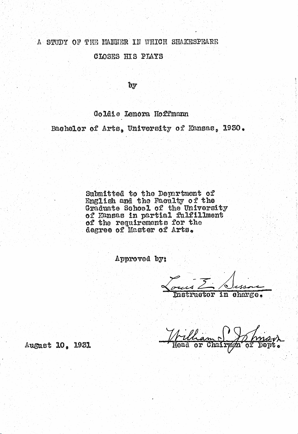
EEG SIGNAL ANALYSIS BEFORE AND AFTER
PERFORMING SALAT ON GAMMA BAND
FARID SHIMAN
RESEARCH REPORT SUBMITTED IN PARTIAL FULFILLMENT FOR THE
DEGREE OF MASTER OF ENGINEERING (BIOMEDICAL)
FACULTY OF ENGINEERING
UNIVERSITY OF MALAYA
KUALA LUMPUR
2012
i
ABSTRAK
Meditasi kerohanian dan upacara penyembahan dilihat sebagai perkara yang
diperlukan untuk keadaan rehat, gaya hidup yang sihat, dan sebagai terapi perubatan
alternatif untuk keseimbangan minda dan tubuh.
Dalam kajian ini, penyelidikan telah dilakukan terhadap semua kesan
neuropsikofisiologik sebelum dan selepas upacara sembahyang dalam Islam (solat)
menggunakan alat elektroensefalogram (EEG). Data EEG dalam kajian ini direkodkan
untuk sepuluh lelaki yang sihat sebelum dan selepas melakukan solat. Untuk menganalisis
data dari isyarat EEG dalam kajian ini, perisian AcqKnowledge 4.0 (BIOPAC Systems Inc,
Goleta, CA) dan Matlab digunakan untuk menentukan dan menganalisis data ketumpatan
spektra kuasa (PSD) (dalam µv²) dan untuk kumpulan sinar Gamma (30-60 Hz). Keputusan
dari kajian ini menunjukkan kuasa sinar gamma dari bacaan EEG mempunyai peningkatan
yang signifikan untuk keadaan selepas solat jika dibandingkan dengan keadaan
sebelumnya. Analisis statistik (paired t-test) menunjukkan ada peningkatan yang signifikan
pada kuasa gamma di bahagian frontal dan occipital otak.
Tambahan lagi, meditasi dalam salat dalam bentuk Perhatian Fokus (FA) dan
Meditasi Transcendetional (TM) telah pertama kali diperkenalkan sebagai proses kognitif
dan corak EEG dalam kajian ini. Kajian ini telah menunjukkan ada perubahan fisiologi
semasa meditasi yang mencadangkan ada tempoh hypometabolik sedar yang mempunyai
kualiti dalam menurunkan aktiviti saraf simpatetik dan meningkatkan aktiviti
parasimpatetik.
ii
ABSTRACT
Religious meditations and prayers were seen as the conditions necessary for
promoting relaxation, healthy living, and acting as alternative medical therapies for
balancing human mind and body. In this study, the investigation of all the
neuropsychophysiological effects of pre- and post-baseline of an Islamic prayer (Salat) on
the electroencephalogram (EEG) was carried out. The EEG data in this study were recorded
for ten healthy males for pre- and post-baseline in the performance of the Salat. In order to
analyze the data from the EEG signals of this study, AcqKnowledge 4.0 software (BIOPAC
Systems Inc, Goleta, CA) and Matlab were used to compute and analyze the power spectral
density (PSD) data (in µv²) for the Gamma (30-60 Hz) band. The results show that the
gamma EEG power has significant increase in post-baseline compared to the pre-baseline.
The statistical analysis (paired t-test) indicated that there was significant increase of gamma
power in the frontal and occipital channels.Additionally, meditation in Salat in the forms of
Focus Attention (FA) and Transcendetional Meditation(TM) were introduced for the first
time as cognitive processes and EEG pattern in this study. This study further revealed that
there are physiological changes during meditation which in turn suggested that there is
wakeful hypometabolic state which has the qualities of decreasing the sympathetic nervous
activity but increasing parasympathetic activity.
iii
ACKNOWLEDGEMENTS
I would like to thank my supervisor, Prof. Fatimah Ibrahim, for her precious guidance,
support, patience, and tolerance throughout the period of this project.
I would like to thank Dr. Ng. Siew Cheok for his comments and support.
I would like to thank Mohammad Javad Safari, Seyed Hossein Safavi, Maysam
Oladazimi, and Fatemeh Molae.
Warmest gratitude to my parents for their love and patience.
iv
TABLE OF CONTENTS
ABSTRAK ............................................................................................................................ i
ABSTRACT ........................................................................................................................ ii
ACKNOWLEDGEMENTS ................................................................................................ iii
TABLE OF CONTENTS ................................................................................................... iv
LIST OF FIGURES ............................................................................................................ vi
LIST OF TABLES ............................................................................................................. vii
LIST OF ABBREVIATIONS ........................................................................................... viii
CHAPTER I: INTRODUCTION ....................................................................................... 1
1.1Overview ............................................................................................................... 2
1.2Problem Statement................................................................................................. 2
1.3Objectives of Study ............................................................................................... 3
1.4Scope of the Study ................................................................................................. 3
1.5Significance of the Study ...................................................................................... 3
1.6Outline of the Report ............................................................................................. 4
2Chapter II: LITERATURE REVIEW ........................................................................... 5
2.1Introduction ........................................................................................................... 5
2.2 Physiology of the Brain…………………………………………………………..5
2.3Electroencephalogram (EEG) ................................................................................ 6
2.3.1Brain Oscillations ............................................................................................. 7
2.4Meditation ............................................................................................................. 8
2.5Salat ..................................................................................................................... 11
2.6Physiological Effects(Autonomic System).......................................................... 13
2.6.1Neurophysiological Effects ............................................................................ 15
2.7Signal Processing................................................................................................. 16
3CHAPTER III: METHODOLOGY ............................................................................ 18
3.1Introduction ......................................................................................................... 18
3.2Subjects ............................................................................................................... 18
3.3EEG Recording and Protocol .............................................................................. 18
3.4Data Analysis....................................................................................................... 19
3.4.1Filter ............................................................................................................... 19
3.4.1.1 Comparison between FIR and IIR………………………………………19
3.4.1.2 IIR filter (Butterworth)…………………………………………………..20
3.4.2Power Spectral Density (PSD) ....................................................................... 21
3.4.3Welch Method ................................................................................................ 21
v
3.4.4 Wavelet Transform……………………………………………………........22
3.5Statistical Analysis .............................................................................................. 24
4CHAPTER IV: RESULTS AND DISCUSSION ....................................................... 25
4.1Introduction ......................................................................................................... 25
4.2 Gamma power in the pre-baseline and post-baseline………………………......25
5CHAPTER V: CONCLUSIONS and FUTURE WORK ............................................ 29
5.1CONCLUSION ................................................................................................... 29
5.2 FUTURE WORK………………………………………………………….……29
6 REFRENCES………………………………………………………………………..30
vi
LIST OF FIGURES
Figure 2.1: Lobes of the brain ..................................................................................... 5
Figure 2.2: Effects of different meditation categories on gamma frequency band... 11
Figure 2.3: Different Positions of performing Salat. ................................................ 12
Figure 2.4: Physiological effects of meditation on the brain .................................... 14
Figure 3.1: Electrodes positions on the scalp………………………………………18
Figure 3.2: Block diagram of FIR and IIR filter ....................................................... 19
Figure 3.3: Comparison between Butterworth, chebyshev and elliptic filters..........20
Figure 4.1: Mean gamma power in pre- and post-baseline in each channel……….27
vii
LIST OF TABLES
Table 4.1: Mean, standard deviation and standard error for gamma power
(B=pre-baseline, A=post-baseline)……………………………….………..26
Table 4.2: Mean and standard deviation (M±S.D) for gamma power in pre- and
post-baseline…………………………………………………………….….28
viii
LIST OF ABBREVIATIONS
EEG Electroencephalography
PSD Power spectral density
SPSS Statistical Package for the Social Sciences
FIR Finite impulse response
IIR Infinite impulse response
Hz Hertz
FA Focus attention
TM Transcendental Meditation
OM Open monitoring
ix
1
1 CHAPTER I: INTRODUCTION
1.1 Overview
The signals from the electroencephalograph (EEG) are seen as the reflections of
neural activities which are recorded in different ways by multiple electrode EEG devices
from inside the brain, under the skull over the cortex, and in certain locations over the scalp
(Sanei & Chambers, 2007). However, EEG was discovered to be an important clinical
device in the field of clinical neurology for diagnosing and monitoring the nervous system
based on the fact that it has the capability to reveal both the normal and abnormal electrical
activity of the brain (Cooper et al., 1980; Niedermeyer & Da Silva, 2005).
There are five major brain waves identified which can be distinguished by their
frequency ranges. These brain waves include the delta (represented by δ) with the
frequency range of between 0.5–4 Hz, theta (represented by θ) with the frequency range of
between 4–8 Hz, alpha (represented by α) with the frequency range of between 8–13 Hz,
beta (represented by β) with the frequency range of between 13–30 Hz ,and gamma
(represented by ϒ) with the frequency range of between 30–128 Hz (Sanei et al., 2007).
Each gamma activity falls above traditional EEG frequency bands which was commonly
considered to range between 30 and 80 Hz. Recently, researches conducted on EEG seemed
to focus more on the increase of the activities in the gamma band for experimental subjects
under meditation because of its relationship with cognitive functioning (Engel et al., 2001;
Fell et al., 2003).
Another study on meditation maintained that it is a body and mind activity which
effects self-regulating practice as it concentrates on training attention for voluntarily
controlling the mental processes (Davidson & Lutz, 2008). The Muslim prayer which is
2
known as Salat in Arabic could be viewed as a form of meditation. This present research
specifically investigates the neurophysiological effects of the Salat as a form of prayer
meditation. This research further unveils the meditation categories in terms of
psychological and neurophysiological effects to achieve better understanding of Salat and
its similarities.
From the frequency plots of EEG signals, it was easily observed that there are some
frequency components up to the range of 25-30 Hz but nothing can be deduced from the
frequencies above 30 Hz. As a result, analysis of gamma waves is not significant from the
frequency plot and a time-frequency approach is the preferred method (Chawla et al.,
2004).
1.2 Problem Statement
Though there is a growing scientific interest in Salat, yet its underlying
neurophysiological mechanism is still under uncertainty. This research therefore
investigates the Salat effects which is a form of meditation. It was observed the benefit of
using Salat is encouraging, developing concentration, clarity and emotional positivity. This
research tends to modify the power spectral density (PSD) in order to make it more robust
for EEG signal processing during performing Salat to extract the features for analyzing the
neurophysiology effects in gamma band (30-60Hz).
1.3 Objectives of Study
The objectives of the study include the following:
i. to compare the two situation of brain activity including the pre- and post-baseline of
the spiritual feeling on Muslim prayer in terms of psychology and neurophysiology
effects.
3
ii. to classify the Salat in Focus Attention (FA) meditation and Transcendental
Meditation (TM) in terms of psychology.
1.4 Scope of the Study
The research is conducted under the following scopes:
i. Development of the method for EEG signals analysis in gamma band.
ii. Investigation of the psycho-physiological effects of Salat meditation for pre- and
post-baseline of the Salat in gamma band.
iii. Determination whether the total power in gamma band after performing Salat (post-
baseline) is higher than the pre-baseline.
iv. Analyzing the EEG signals of subjects in pre- and post-performance of the Salat to
compute the power spectral density (PSD) by using Fast Fourier Transform (FFT)
for total power.
1.5 Significance of the Study
The neurophysiological bases of meditation have been investigated widely in
literature when the activity of meditation produces positive psychological effect both
during and beyond the meditation. In this research, it was illustrated that the EEG signal of
the people who produce spiritual feeling is categorized by an EEG oscillation and their
temporal behavior.
The categorization of the Salat meditation emphasized on the combination of FA
and TM meditation because there is no study to compare the psychophysiology effects
before and after performing Salat meditation but there was an analysis on the EEG signals
in gamma band for mentioned situation. Because high amplitude gamma activity is related
to the cognitive functioning (Barry et al., 2010),current EEG investigation has seen an
increased focus on activity in the gamma band.
4
1.6 Outline of the Report
This research is divided into five chapters and each of the chapters illustrating one
aspect of the project. In Chapter I of this research, introduction and overview of the project
on the subject, statement of the problem, objectives, and significant of the study are
discussed. Chapter II presents an overview of the Electroencephalogram (EEG) and was
followed by the characteristics of Salat meditation. The chapter also reviews the past
literature on the effects of meditation in terms of physiology and neurophysiology.
Chapter III of this study discusses the methodology which is related to the methods,
design, and details of signal analysis used in this research. Chapter IV discusses the result
and discussion about comparison between the pre- and post-effects of Salat meditation in
gamma band in terms of EEG analysis and psychological effects. Finally, chapter V
proposes the conclusion of this study and discusses further work for the research.

5
2 Chapter II: LITERATUREREVIEW
2.1 Introduction
This chapter reviews the literatures regarding physiology of brain, EEG, meditation
and Salat. The literatures on the physiological and neurophysiological effects of meditation
were further reviewed.
2.2 Physiology of Brain
The brain is the majority organ which is a mass of pinkish-gray tissue in the human
body. The brain function is still a mystery for the neurologists with approximately ten
billion neurons. The scientific research of the nervous system and the brain is called
neuroscience or neurobiology (Swanson, 2011). The cortex or cerebrum is the largest part
of the human brain compared to the all parts of the brain which is divided into four lobes:
occipital lobe, parietal lobe, frontal lobe, and temporal lobe (Figure 2.1).
Occipital Lobe: The occipital lobes are the smallest lobes which are located in the
back portion of the skull. It is responsible for visual perception system. The functions of the
occipital lobe include visual processing, color recognition and movement.
Figure 2.1: Lobes of the brain(Swanson, 2011).
6
Parietal Lobe: The parietal lobe is related to the sensory information from different
parts of the body. The functions of the parietal lobe consist of information processing,
movement, spatial orientation, perception of touch and temperature.
Frontal Lobe: The frontal lobe is responsible for higher cognitive purposes. The
functions of the frontal lobe consist of reasoning, short term memory, planning, movement,
behavior, sexual urges, emotions, and organizing parts of speech.
Temporal Lobe: The temporal lobes are responsible for auditory processing as a
primary auditory cortex. This lobe is responsible for arrangement of long term memory.
2.3 Electroencephalogram (EEG)
This chapter presents some basic information on neurophysiology which should be
deemed to be necessary for understanding the experiments and further throw more light on
the results described in the subsequent chapters. Contrarily, this chapter focused on the
description of the electroencephalogram especially as it was applied to the study of
meditation and brain oscillations.
The development of EEG was originally initiated to serve as a method for careful
studying of mental processes. The EEG signal is an act of measuring the current-flows
during synaptic excitations of the dendrites of many pyramidal neurons in the cerebral
cortex. The synaptic currents are produced within the dendrites which could make it
possible for the brain cells (neurons) to be activated (Sanei et al., 2007). However, the
measuring of EEG has been deeply studied to thoroughly examine the underlying brain
physiological changes which have mutual relationship with various states of consciousness
during meditation.
The study conducted by Berger (1929) stated that the first recording of brain
electrical activity which was exposed in the brains of rabbits and monkeys were reported by
7
Caton in 1875. However, it was not fully developed in human beings till 1929 when Hans
Berger made the report for the first measurement of brain electrical activity in humans.
2.3.1 Brain Oscillations
As it was earlier mentioned in this research, there are five major brain waves which
were distinguished by their different frequency ranges. These frequency bands range from
low to high frequencies which include the alpha (α), theta (θ), beta (β), delta (δ), and
gamma (γ). The introduction of alpha and beta waves were done by Berger in the study he
conducted in 1929. In the study conducted by Jasper and Andrews (1938), gamma as a
frequency band was used to refer to the waves which were above 30 Hz. The delta rhythm
was introduced in the study conducted by Walter (1936) to denote all frequencies below the
alpha range. The introduction of theta waves was to denote those having frequencies within
the range of 4–7.5 Hz .More so, theta wave was introduced by Walter and Dovey in 1944
(Sterman et al., 1974; Walter & Dovey, 1944).
Delta rhythms (0.5-4 Hz): The delta rhythms are the waves which are primarily
associated with deep sleep and may be present in the waking state.
Theta rhythms (4-7.5 Hz): The term theta which is presumed to be associated with
the thalamic origin possesses the theta waves which appear as consciousness slips towards
drowsiness. Theta waves were further seen to be associated with having access to
unconscious material, creative inspiration and deep meditation. They are enhanced during
sleep and play an important role in childhood. High theta activity is considered abnormal in
the awaking adult state as it has relationship with different brain disorders.
Alpha rhythms (8-13 Hz): The alpha rhythms appear spontaneously in normal
adults when they are awake, under relaxation and inactive mental conditions. The alpha
8
rhythms could be best seen with eyes closed while mostly pronounced in occipital
locations.
Gamma rhythms (30-60 Hz): These could be seen as the regions of high EEG
frequencies and highest levels of attentive processes which are located in the frontal area.
2.4 Meditation
Meditation is the physiological state necessary for reducing metabolic activity which
in turn brings physical and mental relaxation. Meditation has been viewed from the angle of
enhancing psychological balance and emotional stability (Rubia, 2009).This chapter
reviews meditation effects at the physiological, neurophysiological and affective levels as
well as the scientific paradigms used to study these effects.
Meditation is a common term used to refer to different practices for self-regulation
of emotion and attention which is invariably considered in most religious or philosophical
traditions as an experiential practice (Gunaratana, 2002). More so, meditation usually
involves making one’s attention to be concentrated on a particular physical or mental
object. Practitioners who are involved in meditation are instructed to bring their attention
back to the meditative task whenever their minds start wandering about.
The practice of meditation frequently involves altering the states of consciousness
although meditation could arise when there are intensive practices for meditation for
several hours on daily basis. The practitioners of meditation frequently perform daily
meditation for a certain period of time which could range from 15 minutes to several hours.
There were assumptions that different conscious states could result to different
neurophysiological states while neuroscientific approaches towards meditation focuses on
altered sensory, cognitive and self-awareness experiences.
9
Some studies have discovered that the neurophysiological changes which could be
influenced by meditation are of two kinds. The first of these changes occurred when
meditation practices are referred to as state changes. The second part of the changes (trait
changes) which could be built up over months or years (Cahn & Polich, 2006). However,
emphasis has been made on the importance of the study of meditation states for
consciousness as a means of exploring the effects of meditation on the brain.
It was clearly stated in some studies that there are a large number of meditative
practices but based on the fact that self-regulation of attention is one of the components that
is common, it is imperative to classify styles of meditation based on how the attention
processes are directed (Cahn, et al., 2006). In other studies, meditation practices were
divided into two categories. The first category is the focused attention meditation which
involves the voluntary and sustained attention on a chosen object while the second category
is the open monitoring meditation which involves non-reactive monitoring of the moment-
to-moment content of experience (Lutz et al., 2008).Another study suggested the third
category of meditation practice and automatic self-transcending which includes techniques
designed to transcend their own activity (Travis & Shear, 2010).
Focus Attention (Concentration meditation): The focus attention (FA) meditations
involve continuous sustained attention on a selected object. The object of focus could
include a repeated sound or word (mantra), the breath or body sensations as well as the
imagined mental image. Meditation based on focused attention involves narrowing of
awareness so that the mind only contains the object of focus.
Open monitoring (mindfulness meditations): Many studies have observed that in
mindfulness, practitioners are ordered to permit any thought, feeling or sensation to arise in
consciousness as there is maintenance to a non-reactive awareness to some experiences
(Cahn, et al., 2006; Gunaratana, 2002; Kabat‐Zinn, 2003; Lutz, et al., 2008).
10
Meditation practices which involved having one’s attention focused to a specific
object in the experiential field may lead to a higher activity in the beta and gamma bands
including the meditations from Chinese traditions, Buddhist, and Tibetan Buddhist.
However, the open monitoring which is characterized by theta activity (Austin, 2006;
Gyatso & Jinpa, 1995; Lutz, et al., 2008).
Transcendental Meditation (TM): Transcendental meditation is a form of mantra
(prayer meditation) and is presented in Tibetan Buddhism and Hinduism(Braboszcz et al.,
2010). Mantra could be in the form of religious or mystical sound, word or poem that can
be either recited aloud or sub-vocally. Those who meditate mantra are instructed to focus
their full attention on the recitation as they repeat it and also focus their attention on its
meaning. The practices involving mantra meditation are present in all religions and spiritual
traditions as sutras texts are involved in Buddhism (discourse from the Buddha). Another
instance is Muslim practices (Salat) based on the recitation of a prayer phrase which
involves the slow reading of the Quranic phrases.
As a result, we can classify the Salat as a Focus Attention (FA) and Transcendental
Meditation(TM). In Figure 2.2, It is summarized all findings about meditation categories.

11
2.5 Salat
This could be seen as a form of meditation and also a mandatory practice which
must be performed following certain sets of conditions, set of sequence and at certain set of
times in the Muslim prayer (Alwasiti, 2010). Some studies have observed that religious
meditations and prayers were meant to possibly promote relaxation and healthier conditions
suitable for the human mind and body (Lee et al., 2007; Reibel et al., 2001).Salat has been
considered in the researches for a few years.
Doufesh et al. (2011) investigated EEG signal for alpha band in the different
position of the actual and acted Salat in Muslim prayer(Doufesh, et al., 2011). Their results
indicated that a significantly higher alpha power was recorded during the prostration
position. In other researches, for instance, Haider et al.(2010)illustrated that alpha and theta
power did not increase in their studies but the database was small as they collected the
Meditationcategories
FocusAttention(FA)
Highactivityinthebeta
andgamma(30‐50Hz)
TibetanBuddhisttradition:
increasedfrontal‐parietal
gammacoherenceand
powertradition(Davidson,
2004).
Transcendental
Meditation(TM)
Alphaandgammaactivity
Vipassana
meditation:increase
occipitalgamma
power(Cahnetal.,2010).
OpenMonitoring(OM) Thetaactivity
Figure2.2: Effects of different meditation categories on gamma frequency band

12
database from one subject(Alwasiti, 2010).However, the previous studies confirmed the
high alpha activity with a relaxed state .Alwasiti (2010) research is not valuable to make
decision for effects of Salat (Arambula et al., 2001; Lee, et al., 2007; Reibel, et al., 2001).
The practice of Salat is regarded as being important
in Islam because it is deemed to be beneficial both
physically and mentally to the human body. However, the
recently conducted scientific studies revealed that the Salat
brings physical and mental benefits to the body (Ibrahim et
al., 2008). Additionally, there are involvements of the
physical movements of the body to the fundamental part of
the Muslim prayer in the recitation of verses in Quran and
specific supplications. Despite the fact that there must be
verbal words during meditations, the worshipper should
assume certain positions and perform specific movements.
The four main positions and movements involved in the
prayers include standing, bowing, prostrating and sitting in
figure 2.3 (Ibrahim, et al., 2008; Yucel, 2008).
Standing: This involved subjects having to stand upright.
While on the standing positions, they worshippers’ hands
are placed on the top of the other. This is a situation where
the right hand was placed over the left and both hands
placed above the navel.
Bowing: In bowing position, hands are raised until they are
positions
Standing
Bowing
Prostrating
Sitting
Figure2.3: Different Positions of
p
erforming Sala
t
(Doufesh et al., 2011).
13
level with the ears or shoulder. This is followed by a 90-degree bow, with the hand
touching the knees and pressing down so that the back of the body is horizontal.
Prostrating: This is used in Salat and should be as a situation where the worshipper brings
the hands and knees to the floor. Properly executed prostration involves seven parts of the
body (the forehead, both palms, both knees, the ends of both feet with toes bent) coming
into contact with the floor. The procedure involved in prostration includes the situation
where the hands assume the following positions:
i. The hands are kept away from the sides of the body
ii. The elbow is raised off the floor
iii. The forehead and face are placed in between both palms
iv. The fingers are brought close together
Sitting: The sitting posture which occurs between two prostrations involves resting the
buttocks on the left leg thereby cushioning the leg. In the sitting position, both palms rest
on the thighs with the elbows being placed on the thighs too while the fingers were rested
on the edge of the knees. However, the heel would press on the main muscle at the start of
the thigh which is close to the hip joint when the worshiper is in the sitting position.
2.6 Physiological Effect (Autonomic System)
As earlier mentioned, apart from the general relaxation response, the key subjective
experiences in meditation include the reduction of mental activity and the generation of
positive effects. The obvious target of this present study is to explore the clinical effects of
Salat. The most recent researches on the clinical application of meditation effects are still
very much in infancy but there are still some emerging and concrete evidences that
meditation has positive effects on stress-related diseases and on some neuropsychiatric
disorders(Rubia, 2009).

a
p
c
a
c
t
f
a
m
s
m
p
m
r
a
r
The
a
utonomic
n
p
athways i
n
c
omplicatio
n
a
ctions on t
h
c
hanges du
r
t
he charact
e
f
ight and fl
i
a
nd rest (Ca
h
Med
i
m
editation
p
s
tudies that
m
editation
e
p
resent res
e
m
editation
b
r
eviewed h
o
a
nd the righ
t
In a
d
r
espiratory,
activities o
f
n
ervous syst
e
n
Figure 2.
4
n
in its rea
l
h
eir targete
d
r
ing meditat
i
e
ristics of d
i
ght mecha
n
h
n, et al., 2
0
i
tation has s
t
p
ractices a
n
standard d
a
e
xperiences
e
arch there
f
b
ased on th
e
o
w the left
p
t
part intera
c
d
dition, we
heart rate a
n
Figure 2
.
f
the organ
s
e
m as it is c
o
4
. Although
l
ity as the t
w
d
organs or
t
i
on has the
s
ecreasing i
n
n
isms and i
n
0
06; Jevnin
g
t
rong effect
s
n
d maximu
m
a
ily medita
t
thereby fo
s
f
ore focuse
s
e
similaritie
s
p
art of the
b
c
ts with the
s
can prove
n
d blood pr
.
4: Physiolog
s
and visce
r
o
mposed of
this could
w
o compo
n
t
issues. Ma
n
s
uggestion
t
n
sympathet
n
creased pa
r
g
et al., 199
2
s
on various
m
meditatio
n
t
ion practic
e
s
tering hig
h
s
on the d
a
s
which dai
l
b
rain coope
r
s
ympathetic
the effect
o
essure. Res
p
ical effects o
f
r
a in the bo
the sympat
h
be seen as
n
ents are us
u
n
y studies h
a
t
hat wakefu
l
ic nervous
r
asympathet
2
; Rai et al.,
parts of th
e
n
experienc
e
e
s create lo
w
h
arousal st
a
a
ily medita
t
l
y meditatio
n
r
ates more
w
system.
o
f meditati
o
p
iration con
t
f
meditation
o
dy are bei
n
h
etic and pa
r
a schemati
u
ally believ
e
a
ve shown t
h
l
hypometa
b
activity wh
i
ic activity
m
1988; Youn
g
e
body as th
e
e
s. It has
b
w
arousal s
t
a
tes (Cahn,
t
ion more
t
n
have with
w
ith the par
a
o
n with oth
e
t
rol is one
o
o
n the brain
n
g controlle
d
r
asympathet
i
c view but
e
d to have
h
at the phys
i
b
olic state
w
i
ch is impo
r
m
eant for r
e
g
& Taylor,
e
y function
f
b
een stated
t
ates and
m
et al., 20
0
t
han the
m
Salat. Crai
g
a
sympatheti
c
e
r factors i
n
o
f the possi
b
14
d
by the
i
c neural
there is
opposite
i
ological
w
hich has
r
tant for
e
laxation
1998).
f
or daily
in some
m
aximum
0
6). This
m
aximum
g
(2005)
c
system
n
cluding
b
le ways
15
through which meditation acts on autonomic activity. Respiration was observed as one of
the body’s few autonomic functions that can be controlled and in turn affect functioning of
the autonomic nervous system (Badra et al., 2001; Eckberg et al., 1985). Researchers have
shown that breathing can involuntarily slow down during mantra chanting (Bernardi et al.,
2001) breath counting meditation and simple awareness of the breath (Lehrer et al., 1999).
However, it was also revealed that slower respiration rate during meditation practice brings
about changes in cardiovascular activity which in turn corresponds with the increase in
restorative parasympathetic system activities (Saul, 1990).
This increase in parasympathetic activity could also be assessed through the slowing
down of basal heart rate in worshippers indulging in meditation (Pal & Velkumary, 2004).
Studies have shown that slow breathing has significantly been associated with increased
baroreflex sensitivity. It was also observed that blood pressure decreased after meditation
practice by both healthy individuals and hypertension patients (Carlson et al., 2007;
Manikonda et al., 2007). The improvement in the control of blood pressure is usually seen
as the main sign of balance between parasympathetic and sympathetic activity.
Studies have maintained that Sahaja yoga meditation (daily meditation) has been
indicated to reduce parasympathetic activity (Harrison et al., 2004). This include the
reduction in heart related ailments, respiratory and pulse rates, systolic blood pressure and
oxygen metabolism as well as increasing the skin resistance in short- and long-term
practitioners compared to controls (Rai et al., 1988).
2.6.1 Neurophysiological Effects
Many studies on electrophysiological indicated that brain activity plays an important
role in different concentrative meditation techniques. Low frequency rhythms can be used
16
for investigation of attention and working memory and high frequency rhythms is mainly
used for processing of the contents of experience (Razumnikova, 2007).
Gamma bands (30–50 Hz):
Gamma activity is one of the important frequency ranges which reflects short range
connection for local processing of construction and object recognition (Singer, 1999).There
is a direct relation between gamma activity, brain blood flow and rises of synaptic activity
which is important for considering the long term memories (Niessing et al., 2005).
Researchers found that gamma activity is higher in attended stimuli in compare to
unattended stimuli (Jensen et al., 2007).
Lutz et al. (2004) in their research on long term Buddhist meditation found that ratio
of the gamma to slow rhythm during meditation of long term meditators are higher than
control group. This study shows that there is strong correlation (r > 0.6) between relative
gamma power and years of practice (Lutz et al., 2004).
Research on Chinese and Buddhist meditations revealed that gamma activity in the
post-baseline was higher than per-baseline while alpha activity after doing these
meditations decreased to near zero (Litscher et al., 2001).
Lehmann et al. (2001) researched on five meditation patterns. This study revealed
that center of the gamma (35-44 Hz) gravity is related to meditation methods. Gamma
power at right posterior of the brain changes during visualization and gamma power at left
central of brain changes during verbalization (Lehmann et al., 2001).
2.7 Signal Processing
In studying the EEG signal processing, the literatures of many studies concentrated
on the application of Fourier transform. In addition, it was revealed that the most common
form of analyzing EEG signals is through analyzing these signals in time domain which
17
invariably means direct reading of the potentials coming out from the brain in an
amplitude-time plot (Fisch & Spehlmann, 1999). While as earlier mentioned in this present
research, another way of analyzing EEG signal is through frequency domain which in turn
means viewing its Fourier transform. In addition, the Fourier Transform is computationally
very attractive when it will be calculated by using an efficient algorithm called the Fast
Fourier Transform (Cooley & Tukey, 1965) .
However, analyzing EEG signals with the help of wavelets is of great utility unlike
the Fourier transform where the frequency components cannot be localized. The wavelet
transform is used to dictate when the frequencies are present, determine the duration and
the things that are of great importance to the neurologist. More so, wavelet transforms have
been used for studying EEG signals in different ways whereas the Gabor transforms have
been used to find time-varying spectra (Makeig, 1993). Similarly, some studies have
revealed that discrete wavelets were used to get better time-frequency representation
(Bertrand et al., 2001).

18
3 CHAPTER III: METHODOLOGY
3.1 Introduction
This chapter illustrates the design and implementation, methodology and data
analysis in the research.
3.2 Subjects
The databases collected in this research were selected from ten male Muslim students
in the Medical Informatics Laboratory, University of Malaya (average age 24 years).
Participants were asked to fill the informed consent form and procedures of EEG recording
were explained to them. All subjects were free from neurological and psychiatric disorders.
3.3 EEG Recording and protocol
The EEG signals were collected by eight
AgCl electrodes on the scalp at a sampling
frequency of 500Hz according to the International
10-20 Placement System. The electrodes were
located at frontal (F3, F4), central (C3, C4),parietal
(P3, P4), occipital (O1, O2), and reference electrode
and electrical ground placed at the vertex (Cz) and ear
lobe electrode, respectively (Figure 3.1).
The EEG was collected by eight Biopac EEG100C amplifiers (BIOPAC Systems
Inc., California, USA) .In addition, the output of amplifiers analyzed with the Biopac
MP150 acquisition system. The signals were collected at a sampling frequency of 500Hz.
Figure 3.1: Electrode positions on the scalp

19
The subjects lied on the bed to be relaxed in the supine position for 15 minutes. The
first pre-baseline data were collected with the eyes open for a minute and eye closed for a
minute. Furthermore, EEG signal were collected as a post-baseline after the subjects
perform the Salat, similar in procedure to the pre-baseline data collection.
3.4 Data Analysis
The EEG signals were analyzed by AcqKnowledge 4.0 software (BIOPAC Systems Inc,
Goleta, CA).The data was filtered using (IIR) Butterworth band-pass filter between 1 and
100 Hz. Furthermore, a digital Fast Fourier Transform based on power spectrum analysis
(Welch technique, Hanning windowing function) compute the total power of EEG rhythms
with 60sec epoch for pre- and post-baseline in open eye state for each subjects. The total
power spectral density (PSD) were calculated (in µv²) for the gamma (30-60 Hz) band.
3.4.1 Filter
3.4.1.1 Comparison between FIR and IIR
The filter algorithms are related to infinite impulse-response (IIR)filters which were
designed in analog filters .In this way, the algorithm were developed with several real
coefficients for resulting in digital filter.IIR filters have the feedback in their algorithms in
contrast with FIR filters(Lutovac et al., 2001). Because of feedback, IIR filters have
considerably better frequency response than FIR filters of the same order (Fig 3.2).
Figure3.2: Block diagram of FIR and IIR filter

3
C
f
i
T
w
e
b
o
3
.4.1.2 IIR
f
In th
i
C
hebyshev,
The
f
requency t
h
Butt
e
i
ndicates th
e
T
hese filter
w
hich requi
r
The
a
e
xcellent at
b
an
d
-pass S
o
r offline d
u
f
ilter (Butt
e
i
s section,
w
and elliptic
Butterwort
h
h
an the Che
b
e
rworth filt
e
e
rolls off t
o
s are widel
y
r
ed an suita
b
a
dvantage o
simulating
econd or
d
e
r
u
ring EEG r
e
Figure 3.3:
e
rworth)
w
e compare
a
approximat
i
h
filter roll
s
b
yshev filter
e
rs were de
v
o
wards zero
y
used in t
b
le transfor
m
f the Butter
w
the ban
d
p
a
r
Butterwort
h
e
cordings a
n
Comparison
b
a
nalog filter
i
ons.
s
off more
or the Ellip
t
v
eloped wit
h
in the ban
d
he continu
o
m
ation.
w
orth is rel
a
a
ss of an id
e
h
filters as
a
n
d analysis(
L
b
etween Buter
w
approximat
i
slowly wi
t
t
ic filte
r
(Fi
g
h
cut-off fr
e
d
stop and
m
o
us-time do
m
a
ted to the
f
e
al filter. W
a
cutoff freq
u
L
utovac, et
a
w
orth, chebys
h
i
on includin
g
t
hout rippl
e
g
3.3).
e
quency. T
h
m
aximally f
l
m
ain and d
i
f
lat band pa
s
e present si
m
u
ency that c
al
., 2001).
h
ev and ellipti
c
g
Butterwor
t
e
around th
h
e Butterwo
r
l
at in the p
a
i
screte-time
s
s meaning
m
ple algori
t
an be appli
e
c
filters
20
t
h,
e cutoff
r
th filter
a
ss band.
domain
that it is
t
hms for
e
d online
21
3.4.2 Power Spectral Density (PSD)
The power spectral density (PSD) function
() is described as the Fourier
transform of the autocorrelation function of a given random process (Hyvarinen, 1999).
ΓΥ()=Γ() 3.1
Consequently, the output PSD function is defined by the input PSD function
multiplied by the squared magnitude response of the linear system. This equivalent is
frequency domain description for random signals which is related to the input–output
relationship of a linear time domain system.
Power spectra indicate the distribution of signal’s power for each frequency bands.
This can be computed by square of FFT’s magnitude. When there are not accessible more
data points for each FFT calculation, the frequency resolution (tradeoff) will be
decreased(Kay, 1998).
The spectral windowing increased the accuracy of power spectral density for each
segment is windowed. However, the windowing reduces the contribution of the signal near
the end of segment.
3.4.3 Welch Method
The Welch periodogram can be applied same as the Bartlett periodogram. Similar to
Bartlett's method, the Welch computes the sample of length N by dividing a long sequence
of samples into a set of shorter segments. Moreover, the shorter segments can be applied to
their neighbors for some segment of their length(Kay, 1998). Before each spectrum of
segment’s sample is calculated, w[n] as a data window is concerned to each segment.

22
Advantages of Welch method:
1. The variance of the random process decreases in comparison to basic periodogram and
Bartlett method in Welch methods.
2. All the windowing techniques can apply to the Welch method.
Welch periodogram are computed by four specific steps.
a. Divide the accessible sample sequence into P overlapping segments of D samples each,
with a shift of S<D samples between consecutive segments. If the original sequence is
x[k],the pth segment can be expressed as
3.2
b. W[n] performas data windowto each segment:
0,1, … , 1 3.3
c. Compute discrete frequency sample spectrum for each of the P windowed segments:
0,1, … , 1 3.4
Where
∑|
|
3.5
d. Compute the arithmetic average of the P different sample spectra at each frequency:
∑
0,1, … , 1 3.6
The result, Sm, is the Welch periodogram(Sanei, et al., 2007).
3.4.4 Wavelet transforms
The wavelet transform is a suffusion method in the time- frequency domain
(Chawla, et al., 2004) . Due to this characteristic wavelet transform has widely an
application in signal processing field.

23
Average of Wavelet function
2()LR
ψ
∈ is zero
() 0tdt
ψ
+∞
−∞
=
∫ 3.7
Below equation defines continuous wavelet transform (CWT):
1
(,) () ( )
tb
CWT x a b x t dt
a
a
ψψ
+∞
−∞
−
=∫
3.8
Where x(t) is signal and ()t
ψ
is mother wavelet, a and b are scaling parameters.
The oscillatory frequency and wavelet length is presented by scale translation parameter a
parameter b shows the shifting position. Discrete wavelet based on discrete translation
parameter (b) and scaling parameter (a):
/2
,000
() ( )
mm
mn ta atnb
ψψ
−−
=−
3.9
In general, a=a0m, b=nb0a0m, m shows frequency location and n indicates time
location(Daubechies, 2006).
The wavelet function is obtained by high pass filter and scaling function is obtained
by low pass filter. The decomposition process is started by passing the signal through these
filters, which divide the signal into details (high frequency components) and approximation
(low frequency components). Outputs of filters are decimated by two for obtaining the
approximation coefficients and detail coefficients in the first level (A1, D1). In the next
level approximation coefficients are divided again into approximation and detail
coefficients (A2, D2) until we reach to expected level(Sanei, et al., 2007).
Parseval’s theorem shows that energy of the signal can be classified at different
levels of resolution(Chawla, et al., 2004).
2
1
, 1,...,
N
iij
j
E
DDil
=
==
∑ 3.10

24
2
1
, 1,...,
N
iij
j
E
AAil
=
==
∑ 3.11
Where level of the decomposition indicated by i (from 1 to l) and N is number of
coefficient. EAi and EDi are energies of approximation at detail at decomposition level I
respectively.
3.5 Statistical Analysis
The statistical analysis was used by SPSS to test the gamma power before and after
Salat .In this study, we focus on the two groups of variable in the same subjects. For this
reason, we applied the paired sample t-test to compare the relationship between the pre- and
post-baseline.
The paired sample t-test compares the means of two variables. If the value of
significance is less than 0.05, there is a significant difference. In this research, we applied
the paired sample t-test to compare the mean gamma power in supine position for pre- and
post-baseline.
25
4 CHAPTER IV: RESULTS AND DISCUSSION
4.1 Introduction
In this chapter, the results of gamma power are presented for pre- and post-baseline
in ten subjects. Gamma activity were illustrated when subjects created a positive emotion or
continuous attention on a selected object .In addition, the object of focus maybe on an area
of the body or a repeated word(Quranic phrases).The previous researches investigated the
increase of gamma power in the other prayer meditation(Cahn et al., 2010; Davidson, et al.,
2008).Salat has also been demonstrated as a religious meditation in terms of focus attention
(FA) and Transcendental meditation (TM).
4.2 Gamma power in the pre-baseline and post-baseline
EEG signal of ten subjects (all male) were analyzed for Pre-baseline (before) and
Post-baseline (after).The Paired t-test analysis was performed in comparing measurements.
Table 4.1 indicates the mean, standard deviation, and standard error of gamma power in
each channel for pre- and post-baseline.

26
Mean Std.
Deviation
Std. Error
Mean
O1B .000513565 .0004486682 .0001158456
O1A .000781224 .0005923955 .0001529559
O2B .000466239 .0003490482 .0000901239
O2A .000645481 .0004906886 .0001266953
P3B .000296574 .0002889243 .0000745999
P3A .000719395 .0018581719 .0004797779
P4B .000597583 .0005374781 .0001387762
P4A .000570764 .0006445130 .0001664125
C3B .000234754 .0001970740 .0000508843
C3A .000418015 .0005065278 .0001307849
C4B .000191529 .0002013935 .0000519996
C4A .000218782 .0001725980 .0000445646
F3B .000336198 .0002242707 .0000579064
F3A .000418768 .0003094152 .0000798907
F4B .000268992 .0001428525 .0000368844
F4A .000726245 .0007074690 .0001826677
In addition, Table 4.1 shows that the gamma power in the post-baseline is higher
compared to the pre-baseline of the Salat. The finding is reliable with previous studies on
the neurophysiological correlation that reported an increase in gamma power after
meditation (Davidson, et al., 2008; Travis, et al., 2010). To be more specific, the research
also indicates the difference between right and left hemisphere in pre- and post-baseline of
performing the Salat. The comparison between pre- and post-baseline will be described in
terms of autonomic system (Fig.4.1).
Table 4.1: Mean, standard deviation and standard error for gamma
power (B=pre-baseline, A=post-baseline).

27
Fig 4.1 shows the comparison of mean gamma power at eight channels between pre-
and post-baseline. The channels in the left side of the brain (O1, P3 and C3 except F3) have
higher gamma power than the channels in the right region (O2, P4 and C4 except F4) after
performing the Salat (Post-baseline).
As we mentioned, many studies have shown that the physiological changes during
meditation indicate the effects on autonomic system. The wakeful hypometabolic state has
the characteristics of decreasing in sympathetic activity which is significant for fight and
flight mechanisms and increased parasympathetic nervous activity meant for rest and
relaxation (Cahn, et al., 2006; Rai, et al., 1988). Craig (2005) also indicated how the left
part of the brain cooperates more with the parasympathetic system and the right part
interact with the sympathetic system. Furthermore, we can confirm our finding that the
gamma power will be increased in the left side of the brain during performing the Salat. For
0
0.0001
0.0002
0.0003
0.0004
0.0005
0.0006
0.0007
0.0008
0.0009
O1 O2 P3 P4 C3 C4 F3 F4
POWER V2/Hz
ELECTROD'S LOCATIONS
MEAN GAMMA POWER IN PRE- AND POST-BASELINES
PRE-BASELINE
POST-BASELINE
Figure 4.1: Mean gamma power in pre- and pos
t
-
b
aseline in each channel

28
this reason, we prove the positive effects of the Salat on the parasympathetic nervous
system.
The differences between pre- and post-baseline should be investigated by statistical
analysis methods. As mentioned in methodology, the paired t-test were used to detect the
significant of differences for probability value of sig<0.05.
Paired Differences
t
Sig. (2-
tailed)
Before(pre-
baseline)
After(post
-baseline)
Std.
Error
Mean
95% Confidence Interval of the
Difference
Lower Upper
O1 .0005135±.000448 .0007812±
.0005923
.000099 -.0004804853 -.0000548331 -2.697 .017
O2 .0004662±.0003490 .0006454±
.0004906
.000045 -.0002778483 -.0000806357 -3.899 .002
P3 .0002965± .0002889 .0007193±
.0001858
.000488 -.0020434147 .0000515230 -2.039 .061
P4 .0005975±.0005374 .0005707±
.0006445
.000109 -.0002086544 .0002622932 .244 .811
C3 .0002347±.0001970 .0004180±
.0005065
.000080 -.0003555865 -.0000109359 -2.281 .039
C4 .0001915±.0002013 .0002187±
.0001725
.000013 -.0000569681 .0000024620 -1.967 .069
F3 .0003361±.0002242 .0004187±
.0003094
.000036 -.0001600605 -.0000050791 -2.285 .038
F4 .0002689±.0001428 .0007262±
.0007074
.000155 -.0007913923 -.0001231133 -2.935 .011
Table 4.2 shows the mean, standard deviation, standard error, confidence interval
and P-value of the statistical analysis. There is a significant increase in gamma power in
occipital and frontal regions and left side of the central region (C3). Previous researches
have demonstrated the effects of vipassana and Tibetan Buddhist meditation in the frontal
and occipital regions of the brain(Cahn, et al., 2010; Davidson, 2004). These results have
proved that performing Salat indicates the positive emotion on prayers mind. In addition,
there is no significant difference in the parietal and right side of the central regions as the
probability value is more than 0.05.
Table4.2: Mean and standard deviation (M±S.D) for gamma power in pre- and post-baseline
29
5 Chapter V: CONCLUSIONS AND FUTURE WORK
5.1 CONCLUSION
In conclusion, Salat indicates that the gamma power in the post-baseline is higher
compared to the pre-baseline. The higher gamma activity was described when subjects
performing a continuous attention or positive emotion after performing other prayer
meditation.
This study also showed that the mean gamma power on the left hemisphere is higher
than in the right. Previous studies indicated that increase activity of the left part of the brain
will be increased the parasympathetic system (Craig, 2005).Furthermore, we can prove our
finding that the parasympathetic activity increases after performing the Salat in terms of
physiological effects on the brain. This gamma power difference on the left-side
hemisphere is related to increase of relaxation and a decrease of anxiety.
In addition, there is a significant increase in the occipital and frontal lobes during
the post-baseline of the Salat which is correlated with other researches on vipassana and
Tibetan Buddhist meditation (Cahn, et al., 2010; Davidson, 2004).
5.2 FUTURE WORK
The results of this study can be improved with other bio-potential signals for heart
rate and blood pressure in terms of physiological effects. The research can be extended by
analyzing the EEG signal during performing the Salat to compare it with the pre- and post-
baselines. Finally, by using more subjects, more reliable results will be obtained.
30
REFERENCES
Alwasiti, H. H. EEG activity in Muslim prayer: A pilot study. Maejo International Journal
of Science and Technology, 4.
Arambula, P., Peper, E., Kawakami, M., & Gibney, K. H. (2001). The physiological
correlates of Kundalini Yoga meditation: a study of a yoga master. Applied
Psychophysiology and Biofeedback, 26(2), 147-153.
Austin, J. H. (2006). Zen-brain reflections: Reviewing recent developments in meditation
and states of consciousness: The MIT Press.
Badra, L. J., Cooke, W. H., Hoag, J. B., Crossman, A. A., Kuusela, T. A., Tahvanainen, K.
U. O., et al. (2001). Respiratory modulation of human autonomic rhythms.
American Journal of Physiology-Heart and Circulatory Physiology, 280(6), H2674-
H2688.
Barry, R. J., Clarke, A. R., Hajos, M., McCarthy, R., Selikowitz, M., & Dupuy, F. E.
(2010). Resting-state EEG gamma activity in children with Attention-
Deficit/Hyperactivity Disorder. Clinical Neurophysiology, 121(11), 1871-1877.
Bernardi, L., Sleight, P., Bandinelli, G., Cencetti, S., Fattorini, L., Wdowczyc-Szulc, J., et
al. (2001). Effect of rosary prayer and yoga mantras on autonomic cardiovascular
rhythms: comparative study. Bmj, 323(7327), 1446-1449.
Bertrand, O., Tallon-Baudry, C., Fischer, C., & Pernier, J. (2001). Object representation
and gamma oscillations.
Braboszcz, C., Hahusseau, S., & Delorme, A. (2010). Meditation and neuroscience: from
basic research to clinical practice. Handbook of Integrative Clinical Psychology,
Psychiatry, and Behavioral Medicine: Perspectives, Practices, and Research.
Springer Publishing Co Inc.
31
Cahn, B. R., Delorme, A., & Polich, J. (2010). Occipital gamma activation during
Vipassana meditation. Cognitive processing, 11(1), 39-56.
Cahn, B. R., & Polich, J. (2006). Meditation states and traits: EEG, ERP, and neuroimaging
studies. Psychological bulletin, 132(2), 180.
Carlson, L. E., Speca, M., Faris, P., & Patel, K. D. (2007). One year pre-post intervention
follow-up of psychological, immune, endocrine and blood pressure outcomes of
mindfulness-based stress reduction (MBSR) in breast and prostate cancer
outpatients. Brain, behavior, and immunity, 21(8), 1038-1049.
Chawla, G., Gupta, A., & Sengar, S. (2004, 11-14 Dec. 2004). A wavelet based detection
and analysis of GAMMA rhythms in EEG signals. Paper presented at the Signal
Processing and Communications, 2004. SPCOM '04. 2004 International Conference
on.
Cooley, J. W., & Tukey, J. W. (1965). An algorithm for the machine calculation of
complex Fourier series. Math. Comput, 19(90), 297-301.
Cooper, R., Osselton, J. W., & Shaw, J. C. (1980). EEG technology: Butterworths London.
Craig, A. (2005). Forebrain emotional asymmetry: a neuroanatomical basis? Trends in
cognitive sciences, 9(12), 566-571.
Daubechies, I. (2006). Ten lectures on wavelets: Society for industrial and applied
mathematics.
Davidson, R. J. (2004). Well-being and affective style: neural substrates and biobehavioural
correlates. PHILOSOPHICAL TRANSACTIONS-ROYAL SOCIETY OF LONDON
SERIES B BIOLOGICAL SCIENCES, 1395-1412.
Davidson, R. J., & Lutz, A. (2008). Buddha's Brain: Neuroplasticity and Meditation [In the
Spotlight]. Signal Processing Magazine, IEEE, 25(1), 176-174.
32
Doufesh, H., Faisal, T., Lim, K. S., & Ibrahim, F. (2011). EEG Spectral Analysis on
Muslim Prayers. Applied Psychophysiology and Biofeedback, 1-8.
Eckberg, D. L., Nerhed, C., & Wallin, B. (1985). Respiratory modulation of muscle
sympathetic and vagal cardiac outflow in man. The Journal of physiology, 365(1),
181.
Engel, A. K., Fries, P., & Singer, W. (2001). Dynamic predictions: oscillations and
synchrony in top-down processing. Nature Reviews Neuroscience, 2(10), 704-716.
Fell, J., Fernandez, G., Klaver, P., Elger, C. E., & Fries, P. (2003). Is synchronized
neuronal gamma activity relevant for selective attention? Brain Research Reviews,
42(3), 265-272.
Fisch, B. J., & Spehlmann, R. (1999). Fisch and Spehlmann's EEG primer: basic principles
of digital and analog EEG: Elsevier Science Health Science div.
Gunaratana, H. (2002). Mindfulness in plain English: Wisdom Pubns.
Gyatso, T., & Jinpa, T. (1995). The world of Tibetan Buddhism: An overview of its
philosophy and practice. Somerville: Wisdom Publications.
Harrison, L. J., Manocha, R., & Rubia, K. (2004). Sahaja yoga meditation as a family
treatment programme for children with attention deficit-hyperactivity disorder.
Clinical Child Psychology and Psychiatry, 9(4), 479-497.
Hyvarinen, A. (1999). Fast and robust fixed-point algorithms for independent component
analysis. Neural Networks, IEEE Transactions on, 10(3), 626-634.
Ibrahim, F., Abas, A. B. W., & Ng, S. C. (2008). Salat: Benefit from Science Perspective:
Department of Biomedical Engineering, Univeristy of Malaya.
Jensen, P. S., Arnold, L. E., Swanson, J. M., Vitiello, B., Abikoff, H. B., Greenhill, L. L., et
al. (2007). 3-year follow-up of the NIMH MTA study. Journal of the American
Academy of Child & Adolescent Psychiatry, 46(8), 989-1002.
33
Jevning, R., Wallace, R., & Beidebach, M. (1992). The physiology of meditation: a review.
A wakeful hypometabolic integrated response. Neuroscience & Biobehavioral
Reviews, 16(3), 415-424.
Kabat‐Zinn, J. (2003). Mindfulness‐Based Interventions in Context: Past, Present, and
Future. Clinical psychology: Science and practice, 10(2), 144-156.
Kay, S. M. (1998). Fundamentals of statistical signal processing, Volume II: Detection
theory. Upper Saddle River (New Jersey), 7.
Lee, S. H., Ahn, S. C., Lee, Y. J., Choi, T. K., Yook, K. H., & Suh, S. Y. (2007).
Effectiveness of a meditation-based stress management program as an adjunct to
pharmacotherapy in patients with anxiety disorder. Journal of Psychosomatic
Research, 62(2), 189-195.
Lehmann, D., Faber, P., Achermann, P., Jeanmonod, D., Gianotti, L. R. R., & Pizzagalli, D.
(2001). Brain sources of EEG gamma frequency during volitionally meditation-
induced, altered states of consciousness, and experience of the self. Psychiatry
Research: Neuroimaging, 108(2), 111-121.
Lehrer, P., Sasaki, Y., & Saito, Y. (1999). Zazen and cardiac variability. Psychosomatic
Medicine, 61(6), 812-821.
Litscher, G., Wenzel, G., Niederwieser, G., & Schwarz, G. (2001). Effects of QiGong on
brain function. Neurological research, 23(5), 501-505.
Lutovac, M. D., Tošić, D. V., & Evans, B. L. (2001). Filter design for signal processing
using MATLAB and Mathematica: Miroslav Lutovac.
Lutz, A., Greischar, L. L., Rawlings, N. B., Ricard, M., & Davidson, R. J. (2004). Long-
term meditators self-induce high-amplitude gamma synchrony during mental
practice. Proceedings of the National Academy of Sciences of the United States of
America, 101(46), 16369.
34
Lutz, A., Slagter, H. A., Dunne, J. D., & Davidson, R. J. (2008). Attention regulation and
monitoring in meditation. Trends in cognitive sciences, 12(4), 163-169.
Makeig, S. (1993). Auditory event-related dynamics of the EEG spectrum and effects of
exposure to tones. Electroencephalography and Clinical Neurophysiology, 86(4),
283-293.
Manikonda, J., Störk, S., Tögel, S., Lobmüller, A., Grünberg, I., Bedel, S., et al. (2007).
Contemplative meditation reduces ambulatory blood pressure and stress-induced
hypertension: a randomized pilot trial. Journal of human hypertension, 22(2), 138-
140.
Niedermeyer, E., & Da Silva, F. H. L. (2005). Electroencephalography: basic principles,
clinical applications, and related fields: Lippincott Williams & Wilkins.
Niessing, J., Ebisch, B., Schmidt, K. E., Niessing, M., Singer, W., & Galuske, R. A. W.
(2005). Hemodynamic signals correlate tightly with synchronized gamma
oscillations. Science, 309(5736), 948.
Pal, G., & Velkumary, S. (2004). Effect of short-term practice of breathing exercises on
autonomic functions in normal human volunteers. The Indian journal of medical
research, 120(2), 115.
Rai, U., Setji, S., & Singh, S. (1988). Some effects of Sahaja Yoga and its role in the
prevention of stress disorders. Journal of International Medical Sciences, 19, 23.
Razumnikova, O. M. (2007). Creativity related cortex activity in the remote associates task.
Brain research bulletin, 73(1-3), 96-102.
Reibel, D. K., Greeson, J. M., Brainard, G. C., & Rosenzweig, S. (2001). Mindfulness-
based stress reduction and health-related quality of life in a heterogeneous patient
population. General Hospital Psychiatry, 23(4), 183-192.
35
Rubia, K. (2009). The neurobiology of meditation and its clinical effectiveness in
psychiatric disorders. Biological psychology, 82(1), 1-11.
Sanei, S., & Chambers, J. (2007). EEG signal processing: Wiley-Interscience.
Saul, J. P. (1990). Beat-to-beat variations of heart rate reflect modulation of cardiac
autonomic outflow. Physiology, 5(1), 32-37.
Sterman, M., Macdonald, L., & Stone, R. K. (1974). Biofeedback training of the
sensorimotor electroencephalogram rhythm in man: effects on epilepsy. Epilepsia,
15(3), 395-416.
Swanson, L. W. (2011). Brain architecture: Understanding the basic plan: Oxford Univ Pr.
Travis, F., & Shear, J. (2010). Focused attention, open monitoring and automatic self-
transcending: Categories to organize meditations from Vedic, Buddhist and Chinese
traditions. Consciousness and cognition, 19(4), 1110-1118.
Walter, W., & Dovey, V. (1944). Electro-encephalography in cases of sub-cortical tumour.
Journal of Neurology, Neurosurgery & Psychiatry, 7(3-4), 57.
Young, J. D. E., & Taylor, E. (1998). Meditation as a voluntary hypometabolic state of
biological estivation. News in physiological sciences, 13, 149-153.
Yucel, S. (2008). The effects of prayer on Muslim patients' well-being. BOSTON
UNIVERSITY SCHOOL OF THEOLOGY.