Influence of biomechanical models on joint kinematics and kinetics in baseball pitching PDF Free Download
1 / 15/15
100%

Delft University of Technology
Influence of biomechanical models on joint kinematics and kinetics in baseball pitching
Gasparutto, Xavier; van der Graaff, Erik; van der Helm, Frans C.T.; Veeger, Dirkjan H.E.J.
DOI
10.1080/14763141.2018.1523453
Publication date
2018
Document Version
Final published version
Published in
Sports Biomechanics
Citation (APA)
Gasparutto, X., van der Graaff, E., van der Helm, F. C. T., & Veeger, D. H. E. J. (2018). Influence of
biomechanical models on joint kinematics and kinetics in baseball pitching.
Sports Biomechanics
.
https://doi.org/10.1080/14763141.2018.1523453
Important note
To cite this publication, please use the final published version (if applicable).
Please check the document version above.
Copyright
Other than for strictly personal use, it is not permitted to download, forward or distribute the text or part of it, without the consent
of the author(s) and/or copyright holder(s), unless the work is under an open content license such as Creative Commons.
Takedown policy
Please contact us and provide details if you believe this document breaches copyrights.
We will remove access to the work immediately and investigate your claim.
This work is downloaded from Delft University of Technology.
For technical reasons the number of authors shown on this cover page is limited to a maximum of 10.

Full Terms & Conditions of access and use can be found at
http://www.tandfonline.com/action/journalInformation?journalCode=rspb20
Sports Biomechanics
ISSN: 1476-3141 (Print) 1752-6116 (Online) Journal homepage: http://www.tandfonline.com/loi/rspb20
Influence of biomechanical models on joint
kinematics and kinetics in baseball pitching
Xavier Gasparutto, Erik van der Graaff, Frans C. T. van der Helm & Dirkjan H.
E. J. Veeger
To cite this article: Xavier Gasparutto, Erik van der Graaff, Frans C. T. van der Helm & Dirkjan H.
E. J. Veeger (2018): Influence of biomechanical models on joint kinematics and kinetics in baseball
pitching, Sports Biomechanics, DOI: 10.1080/14763141.2018.1523453
To link to this article: https://doi.org/10.1080/14763141.2018.1523453
© 2018 The Author(s). Published by Informa
UK Limited, trading as Taylor & Francis
Group.
View supplementary material
Published online: 28 Nov 2018.
Submit your article to this journal
Article views: 312
View Crossmark data

ARTICLE
Influence of biomechanical models on joint kinematics and
kinetics in baseball pitching
Xavier Gasparutto
a
, Erik van der Graaff
a,b
, Frans C. T. van der Helm
a
and Dirkjan H. E. J. Veeger
a,b
a
Department of BioMechanical Engineering, Delft University of Technology, Delft, The Netherlands;
b
Department of Human Movement Sciences, Vrije Universiteit Amsterdam, Amsterdam, The Netherlands
ABSTRACT
In baseball pitching, biomechanical parameters have been linked
to ball velocity and potential injury risk. However, although the
features of a biomechanical model have a significant influence on
the kinematics and kinetics of a motion, this influence have not
been assessed for pitching. The aim of this study was to evaluate
the choice of the trunk and shoulder features, by comparing two
models using the same input. The models differed in thoraco-
humeral joint definition (moving or fixed with the thorax), joint
centre estimation, values of the inertial parameters and computa-
tional framework. One professional pitcher participated in the
study. We found that the different features of the biomechanical
models have a substantial influence on the kinematics and kinetics
of the pitchers. With a fixed thoraco-humeral joint the peak aver-
age thorax angular velocity was delayed and underestimated by
17% and the shoulder internal rotation velocity was overestimated
by 7%. The use of a thoraco-humeral joint fixed to the thorax will
lead to an overestimation of the rotational power at the shoulder
and will neglect the power produced by the forward and upward
translation of the shoulder girdle. These findings have direct
implications for the interpretation of shoulder muscle contribu-
tions to the pitch.
ARTICLE HISTORY
Received 27 July 2017
Accepted 3 September 2018
KEYWORDS
Inverse dynamics;
modelling; overhand throw;
shoulder; trunk
Introduction
Baseball pitching is one of the most studied motions in sport biomechanics, with many
studies aiming to identify the biomechanical variables that influence performance and
the risk of injury (Fortenbaugh, Fleisig, & Andrews, 2009; Oyama, 2012; Weber,
Kontaxis, Brien, & Bedi, 2014; Whiteley, 2007). However, these studies did not all use
the same biomechanical model. Different studies might have important differences in
their biomechanical models, and so produce substantial differences in the estimated
kinematics and kinetics of a given motion.
For a biomechanical model of pitching, we identified four important features (1) the
thoraco-humeral joint model that can be either fixed with the thorax if the thorax is
defined by the hip joint centres and acromio-clavicular joints (Aguinaldo & Chambers,
CONTACT Dirkjan H. E. J. Veeger h.e.j.veeger@tudelft.nl
Supplemental data for this article can be accessed here.
SPORTS BIOMECHANICS
https://doi.org/10.1080/14763141.2018.1523453
© 2018 The Author(s). Published by Informa UK Limited, trading as Taylor & Francis Group.
This is an Open Access article distributed under the terms of the Creative Commons Attribution-NonCommercial-NoDerivatives License
(http://creativecommons.org/licenses/by-nc-nd/4.0/), which permits non-commercial re-use, distribution, and reproduction in any
medium, provided the original work is properly cited, and is not altered, transformed, or built upon in any way.

2009; Fleisig, 1994; Naito, Takagi, & Maruyama, 2011; Roach & Lieberman, 2014)or
moving with respect to the thorax if the thorax is defined by thoracic markers
(Gasparutto, Van Der Graaff, Van Der Helm, & Veeger, 2016; Naito, Takagi,
Yamada, Hashimoto, & Maruyama, 2014; Takagi et al., 2014), (2) the body segment
inertial parameters (Ae, Tang, & Yokoi, 1992; Clauser, Mc Conville, & Young, 1969;
Dempster, 1955; Dumas, Cheze, & Verriest, 2007), (3) the joint centres estimation (Ae
et al., 1992; Dempster, 1955; Dillman, Fleisig, & Andrews, 1993; Dumas et al., 2007) and
(4) the computational framework (Feltner & Dapena, 1986; Gasparutto et al., 2016;
Naito & Maruyama, 2008). In studies of human gait, the features of a biomechanical
model are known to affect the estimation of the kinematics and kinetics of the lower
limb (Dumas, Nicol, & Chèze, 2007; Pearsall & Costigan, 1999; Rao, Amarantini,
Berton, & Favier, 2006; Reinbolt, Haftka, Chmielewski, & Fregly, 2007; Stagni,
Leardini, Cappozzo, Benedetti, & Cappello, 2000). To our knowledge, the influence of
the features of a biomechanical model has not been quantified for baseball pitching, and
so the consistency of the results obtained from the various models used in the literature
has not been verified. It is reasonable to assume that modelling assumptions will also
have a significant influence on the kinematics and kinetics for a highly dynamic motion
as pitching. What is at stake is that significant differences in the estimated kinematics
and kinetics obtained by different models for the same motion could result in contra-
dictory conclusions, specifically for studies using correlations between kinematic and
kinetic parameters. This could lead to incorrect and even potentially harmful recom-
mendations to the coaches and pitchers. In addition, contradictory observations
between two different studies could be due to different modelling assumptions more
than differences in the pitching motion itself. To gain confidence in the recommenda-
tions from the scientificcommunity, it is necessary to understand the influence of the
choice for different features. Therefore, the aim of this study is to evaluate quantitatively
the effect of the trunk and shoulder features by comparing the kinematics and kinetics
obtained by multiple models for the same input pitching motion.
Two different models were selected. The first model was previously developed by the
authors of the present study (Gasparutto et al., 2016) and based on the work of Dumas
et al. (Dumas et al., 2007). The second model was developed by Fleisig et al. (Fleisig, 1994;
Zheng, Fleisig, Barrentine, & Andrews, 2004) and is one of the most used models in
biomechanical studies of baseball pitching. These two models use different regression
equations to determine the inertial parameters and joint centres and have different
mechanical frameworks but the main difference concerns the definition of the thorax
segment and shoulder joint model. The Gasparutto model estimates the thorax motion
with markers on the thorax only whilst the Fleisig model uses the estimated shoulder
joint centres and hip joint centres to represent the thorax. As a consequence the Fleisig
model merges the scapular girdle motion with the thorax motion, whereas, the
Gasparutto model separates between the thorax motion and the displacement of the
scapular girdle relative to the thorax. This leads to the following limitations: (a) if
the thorax is flexed but the shoulders stay above the hips, the Fleisig model will not
capture this flexion, (b) if the scapular girdle is moved forward, the Fleisig model will
interpret that motion as an axial rotation of the thorax. Although this feature is
bound to have an effect on thorax and arm kinematics, its influence was never assessed
previously. Based on the limitations of the Fleisig model, we hypothesise that the merged
2X. GASPARUTTO ET AL.

thoraco-humeral joint model will show reduced thorax flexion and tilt and increased axial
rotation leading to increased shoulder angles, angular velocities and actions when com-
pared to the moving thoraco-humeral joint model developed by Gasparutto et al. (2016).
Methods
One professional right-handed baseball pitcher, with 5 years of MLB experience,
participated in the study (height: 1.98 m, weight: 101.2 kg). After having been informed
of the aims and procedures of the experiment, the player signed an informed consent
form. The Faculty of Human Movement Sciences’local ethical committee approved this
research project.
Equipment
A motion capture system consisting of eight motion capture beams was used to track
active skin markers (4 Optotrack Certus, 4 Optotrack 3020, Northern Digital Inc.,
Waterloo, Ontario, Canada). The pitching mound consisted of a two-part wooden
pitching mound that was taped to two forceplates (Vrije Universiteit, Amsterdam,
The Netherlands,1.08 x 1.08 m, 200 Hz, (Ibrahim, Faber, Kingma, & van Dieën,
2017)). The standing part of the pitching mound included a pitching rubber and the
stepping part had a downward slope of 5.5 degrees as recommended in the Major
League Baseball regulations. A high-speed camera (Casio EX-ZR 1000, Casio Computer
CO., LTD., Tokyo, Japan) was used to film the mound from the right lateral side at a
frame rate of 240 Hz.
A net with a rectangular pitching target was placed at 10 m from the mound. As the
strike zone is at 18 m during a game, the target was scaled to the usual strike zone. The
pitches were marked ‘strike’if they reached the target and ‘ball’if they were out of
the target. A speed gun (Stalker Pro II Speed Sensor Radar, Applied Concepts, Inc./
Stalker Radar, Plano, Texas, United States) placed behind the net was used to record the
maximal ball speed of every pitch in miles per hour (mph).
Measurement procedure
The pitcher had a one-hour warm-up with his physical trainer, as in game conditions.
The pitcher was then equipped with 24 active markers; 18 were placed on anatomical
points on the head, upper limbs and lower limbs and 2 clusters of 3 active markers on
the thorax and pelvis respectively.
Once equipped with markers, the pitcher performed as many warm-up throws as he
wished from the pitching mound. When ready, the pitcher performed from the mound
three fastballs with six active markers on the upper limb and then eight fastballs with 24
active markers on the full body. The acquisition was done at the maximal acquisition
frequency: 170 Hz with six markers and 90 Hz with 24 markers.
The pitcher was instructed to throw as fast as he could while attempting to hit the
pitching target. After the throwing procedure, eight anatomical points of the pelvis and
thorax were acquired with a pointer. The main markers used in this study are depicted
on Figure 1.
SPORTS BIOMECHANICS 3

Data processing
Data processing was done with Matlab2014a (The MathWorks, Inc., Natick,
Massachusetts, United States) and with the use of Mokka and of the BTK Matlab API
(Barre & Armand, 2014). The five fastest strikes were used for the analysis.
Marker trajectories and ground reaction forces
The marker trajectories were interpolated and upsampled to 170 Hz with a validated
upsampling method (see Supplementary Materials). The upsampled marker trajectories
were filtered with a fourth order Butterworth low-pass filter with a cutting frequency of
12.5 Hz during the stride and follow-through and with a cutting frequency of 25 Hz
during the arm cocking, the arm acceleration and the arm deceleration phases.
The thorax and pelvis anatomical points were reconstructed based on the pointing
procedure and the pelvis and thorax cluster motion. The joint centres (shoulder, elbow,
wrist, hip) were defined with regression equations (Dumas, Cheze et al., 2007). To avoid
inconsistencies in segment length due to the soft tissue artefacts and the interpolation of
the trajectories, a quasi-static multibody optimisation (Lu & O’Connor, 1999), detailed
in the Supplementary Materials, was performed with the ‘fmincon’function of
Matlab2014a.
The synchronised ground reaction force was downsampled to 170 Hz and corrected
for the mound weight. The time of foot contact was defined as the time when the
ground reaction force under the stride foot was larger than 10 N.
The synchronised high speed camera was used to identify the time of ball release. It
was defined as the first frame when the ball is not visibly touched by the hand.
Models
The Fleisig model and the Gasparutto model differed in four features: (a) the thoraco-
humeral joint that was fixed or moving, (b) the inertial parameters based on (Dempster,
Figure 1. Markers and optimised length (blue lines).
4X. GASPARUTTO ET AL.

1955) or (Dumas, Cheze et al., 2007), (c) the joint centres estimation based on (Dillman
et al., 1993) or (Dumas, Cheze et al., 2007) and the mechanical framework based on
(Feltner & Dapena, 1986) or (Dumas, Aissaoui, & de Guise, 2004; Gasparutto et al.,
2016). These points are summarised in Table 1 and details of the thoraco-humeral joint
models and mechanical framework are given below.
Thoraco-humeral joint
The Gasparutto model features a ‘moving’thoraco-humeral joint. The thorax is defined
by thorax markers (deepest point of Incisura Jugularis, Processus Xyphoideus,
Processus Spinosus of the 7
th
cervical vertebrae, Processus Spinosus of the 8
th
thoracic
vertebrae, see Figure 1) according to the recommendation of the International Society
of Biomechanics (ISB) (Wu et al., 2005) and the scapular girdle motion is modelled by
allowing the gleno-humeral joint (GH) to translate with respect to the thorax in three
directions (forward/backward, upward/downward, lateral/medial). The Fleisig model
features a ‘fixed’thoraco-humeral joint. The thorax is defined by the right and left hip
joint centres and the right and left GH. The GH displacements are defined with respect
to the midpoint of the right and left GH and are limited to the lateral-medial direction.
A forward/backward motion of the scapular girdle will be interpreted as an axial
rotation of the thorax. Likewise an upward/downward motion of the scapular girdle
will be interpreted as a lateral tilt of the thorax. The geometric differences between the
Gasparutto model and the Fleisig model are depicted in Figure 2.
Kinematics and kinetics
The joint angles represent the angles of the distal segment with respect to the proximal
one and the net joint forces and moments represent the net forces and moments of the
proximal segment on the distal segment. When needed, the sign of the angles from the
Fleisig model and the zero angle position of the joints were modified to match
the recommendation of the ISB and be compatible with the other models. The dis-
placements and velocities of GH in the thorax were computed with respect to the
cervical joint centre for the Gasparutto model and with respect to the mid-point of the
right and left GH for the Fleisig model (1994).
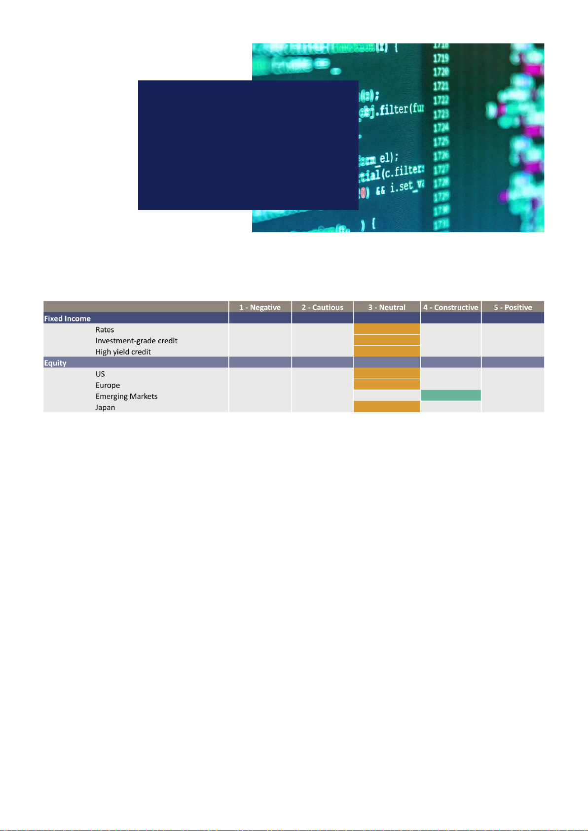
Table 1. Overview of the biomechanical features of the models.
Thoraco-Humeral
Joint
Body Segment Inertial
Parameters Joint Centres Mechanical Framework
Main Models
Fleisig Lumped (Fleisig,
1994)
(Dempster, 1955) (Dillman et al., 1993) (Feltner & Dapena, 1986)
Gasparutto Moving (Gasparutto
et al., 2016)
(Dumas, Cheze et al.,
2007)
(Dumas, Cheze et al.,
2007)
(Dumas et al., 2004;
Gasparutto et al., 2016)
Intermediate Models
Fixed Lumped (Fleisig,
1994)
(Dumas, Cheze et al.,
2007)
(Dumas, Cheze et al.,
2007)
(Dumas et al., 2004;
Gasparutto et al., 2016)
Inertial Moving (Gasparutto
et al., 2016)
(Dempster, 1955) (Dumas, Cheze et al.,
2007)
(Dumas et al., 2004;
Gasparutto et al., 2016)
Joint Centre Moving (Gasparutto
et al., 2016)
(Dumas, Cheze et al.,
2007)
(Dillman et al., 1993) (Dumas et al., 2004;
Gasparutto et al., 2016)
Framework Lumped (Fleisig,
1994)
(Dempster, 1955) (Dillman et al., 1993) (Dumas et al., 2004;
Gasparutto et al., 2016)
SPORTS BIOMECHANICS 5

The joint kinetics were estimated by the inverse dynamics methods described by
Dumas et al. (2004) for the Gasparutto model and by the inverse dynamics method
described by Zheng et al. (2004) for the Fleisig model. The net joint forces and
moments were then projected in the corresponding joint coordinate systems as
explained in Gasparutto et al. (2016).
Intermediate models
To identify the influence of each feature separately, four intermediate models were
built. Three intermediate models were the Gasparutto model with one biomechani-
cal feature of the Fleisig model. The last intermediate model was the Fleisig model
implemented in the mechanical framework of the Gasparutto model. Thus, by
comparing these models to the Gasparutto model we could identify the influence
of each feature separately. The features of the intermediate models are detailed in
Table 1.
The ‘fixed’intermediate model was the Gasparutto model with the thoraco-
humeral joint of the Fleisig model, the ‘inertial’intermediate model was the
Gasparutto model with the inertial parameters from the Fleisig model, the ‘joint’
intermediate model was the Gasparutto model with the joint centres from the Fleisig
model and the ‘framework’intermediate model was the Fleisig model expressed
within the Gasparutto computational framework, that is the Gasparutto model with
the joint centres, inertial parameters and thorax from the Fleisig model. These
definitions are regrouped in Table 1.
Figure 2. Geometric models of the Gasparutto model (blue mesh, spherical markers) and of the
Fleisig model (black, dashed lines, square markers) at ball release.
6X. GASPARUTTO ET AL.
Model comparison
The joint kinematics and joint kinetics estimated by the six models were compared for
parameters relevant to baseball pitching (Fortenbaugh et al., 2009).
The selected kinematic parameters were: the shoulder horizontal abduction angle at
foot contact, the shoulder maximal external rotation angle, the elbow flexion angle at
maximal shoulder external rotation, the peak norm of the thorax angular velocity, the
shoulder peak internal rotation velocity and the elbow peak extension velocity. The
time series of the thorax angles, thorax angular velocities, GH translation and GH linear
velocities were also compared and reported as they represent the main difference
between the two selected models.
Regarding the kinetics, the selected parameters were the peak shoulder pulling force,
the peak of the norm of the elbow net force, the peak of the norm of the shoulder net
force, the peak elbow adduction moment, the peak shoulder external-rotation moment
and the peak shoulder internal-rotation moment.
The mean difference between the parameters from the Gasparutto model and the
Fleisig and intermediate models were computed as well as the timing difference for the
peak values. A two-tailed paired t-test was performed to account for the statistical
significance of the differences.
Results
The mean ball velocity was 39.7 ± 0.2 m/s (88.8 ± 0.4 mph).
GH displacements (Figure 4)
The ‘moving’thoraco-humeral models showed a backward displacement during the
arm cocking phase. This was followed by a forward motion during the arm acceleration
phase with a peak forward velocity of 1.5 m/s at ball release for the Gasparutto model
and a peak forward velocity of 1.9 m/s between ball release and maximal internal
rotation for the ‘joint centre’intermediate model. GH continued with a forward motion
during the follow-through. Regarding the upward motion, the ‘moving’thoraco-hum-
eral models showed small variations in position but an upward velocity during the
acceleration and deceleration phases with a peak of 0.4 m/s and 0.7 m/s at ball release
for the Gasparutto model and ‘joint centre’intermediate model respectively. By defini-
tion, the fixed thoraco-humeral models showed no displacement and no velocity for
these two degrees-of-freedom. All models presented some lateral-medial displacement
and velocity.
Model comparison (Figure 3,Table 2)
When compared to the Gasparutto model the Fleisig model showed increased shoulder
horizontal abduction at foot contact, reduced thorax angular velocity, increased elbow
extension velocity, and increased elbow forces, elbow adduction moment and shoulder
internal rotation moment. The peak thorax angular velocity and peak elbow extension
velocity were also delayed by 15 ms and 11 ms respectively.
SPORTS BIOMECHANICS 7

Based on the ‘fixed’intermediate model results, the thoraco-humeral feature was
found responsible for the reduced thorax angular velocity and showed as well an
increased shoulder internal rotation velocity. The elbow extension velocity was mainly
influenced by the joint centre estimation.
The net joint forces and moments were influenced by the inertial parameters and
the joint centres but the largest influence was found for the mechanical framework
feature.
Figure 3. Thorax angles and angular velocities with respect to the global frame, t = 0 is foot contact
and the vertical lines indicates maximal external rotation, ball release and maximal internal rotation.
Figure 4. Gleno-humeral joint position and velocities with respect to the thorax, t= 0 is foot contact
and the vertical lines indicates maximal external rotation, ball release and maximal internal rotation.
8X. GASPARUTTO ET AL.

Table 2. Mean parameters of the Gasparutto model and differences with the Fleisig model and intermediate models. Only the significant differences were
reported in table (p< 0.05). FC stands for the Foot Contact event, MER stands for the event ‘shoulder Maximal External Rotation’,‘t’is the time at which the
considered parameter occurs, ‘Δt’is the time difference in milliseconds between the models, ‘mean Δ’is the mean difference of the considered parameter.
Angles (deg) Angular Velocity (deg/s) Net Forces (N) Net Moment (Nm)
Shoulder
Horizontal.
Abduction At
FC
Shoulder
Maximal
External
Rotation
Elbow
Flexion
at MER
Peak
Norm of
Thorax
Ang. Vel.
Peak
Shoulder
Internal
Rotation
Peak
Elbow
Extension
Peak
Shoulder
Pulling
Force
Peak Norm
of Elbow
net force
Peak Norm
of Shoulder
net force
Peak
Elbow
Adduction
Peak
Shoulder
Internal
Rotation
Peak
Shoulder
External
Rotation
Results model
Gasparutto
T(ms) 0 204 204 152 249 237 237 236 238 208 208 258
Mean –25 –177 83 1280 3939 –1879 1224 1058 1306 83 83 –36
SD ±9 ±1 ±3 ±21 ±379 ±147 ±24 ±40 ±38 ±1 ±1 ±7
Difference with model Gasparutto
Fleisig Δt(ms) 0 - 0 15 - 11 - –4-33 -
Mean Δ12 - 4 –215 - 118 - 162 - 17 21 -
pvalue 0.006 - 0.000 0.000 - 0.049 - 0.003 - 0.000 0.000 -
Fixed Δt(ms) - 0 - 29 0 - - - - - - -
Mean Δ-5-–165 317 - - - - - - -
pvalue - 0.000 - 0.000 0.000 - - - - - - -
Inertial
Parameters
Δt(ms) - - - - - - 1 3 1 - 3 –1
Mean Δ------–27 –34 –50 - 3 –8
pvalue - - - - - - 0.009 0.000 0.000 - 0.000 0.000
Joint Centre Δt(ms) - - 0 - 0 –2- 4 4 1 3 –2
Mean Δ--–1- 6 83 - 58 –47 2 8 –3
pvalue - - 0.000 - 0.027 0.002 - 0.018 0.029 0.011 0.000 0.008
Mechanical
Framework
Δt(ms) 0 0 0 28 –109 - 6 2 4 –1
Mean Δ13 –4–1 -137 276 77 –43 - –89 3 10 –10
pvalue 0.005 0.003 0.000 0.000 0.000 0.005 0.021 - 0.002 0.002 0.000 0.002
SPORTS BIOMECHANICS 9

Discussion and implications
This study aimed at understanding the influence of the choice of biomechanical model
features on the analysis of pitching, especially for the thorax segment and the thoraco-
humeral joint. Two models were compared: the Fleisig model (Fleisig, 1994; Zheng
et al., 2004) and the Gasparutto model (Gasparutto et al., 2016). The influence of each
feature was evaluated with four intermediate models. Although this paper studied the
pitching motion of a skilled professional baseball player, the possibility of idiosyncrasies
in the pitching technique of the participant cannot be excluded.
This study showed that there is a non-negligible displacement of GH during the
pitch that a fixed thoraco-humeral joint cannot account for. The position of GH with
respect to the thorax showed a backward displacement during the cocking phase
followed by a peak forward and upward velocity occurring at ball release and a forward
displacement during the follow-through, likely to be used to ease the deceleration of the
upper limb.
It is important to note that the lateral-medial displacement of GH does not represent
a shortening-lengthening of the clavicle but the displacement of GH around an arc of
ellipsoid for the moving thoraco-humeral joints and the variation of distance between
the right and left GH for the fixed thoraco-humeral joints. The ‘fixed’model can only
capture lateral-medial displacement of the scapular girdle. However, the lateral/medial
position of GH for the fixed thoraco-humeral joint convention is equal to half the
distance between the left and right GH and the point of reference to compute that
position is moving with respect to the thorax which makes any interpretation difficult.
Thus the fixed thoraco-humeral convention is not appropriate for the description of the
scapular girdle motion and while not exactly modelling the role of the scapula, a
moving thoraco-humeral joint should be preferred to get insight in the scapular girdle
motion during pitching.
The assumption in the Fleisig model that the thorax is defined based on the shoulder
joint centres and hip joint centres led to reduced thorax flexion angle, tilt angle, and
peak thorax rotation velocities and consequently to an increased estimation of the peak
shoulder internal rotation velocity. The increased thorax axial rotation after ball release
was likely to be due to the forward motion of GH during the arm acceleration phase
and follow-through. Indeed, the forward motion of the right GH was interpreted as
thorax axial rotation by the fixed thoraco-humeral joint model. It is interesting to see
that the timing of the peak thorax angular velocity and of the peak elbow extension
velocity was changed. This could lead to significant differences when studying the
kinetic chain. Regarding the kinetics, contrary to our initial hypothesis, the thoraco-
humeral joint definition did not have any significant influence on the net joint forces
and moments. This can be understood by the fact that the equations used to estimate
the net moment and force at the shoulder only use the estimation of the GH position in
the mound reference frame and not the thorax position and orientation. The thorax
orientation was only used during the projection of the shoulder net joint moment and
forces in the shoulder joint coordinate system.
Asignificant difference was found for the peak shoulder internal rotation velocity
between the Gasparutto model and the intermediate models with the fixed thoraco-
humeral joint but not between the Gasparutto model and the Fleisig model. The
10 X. GASPARUTTO ET AL.

intermediate models results suggest that this inconsistency comes from the differences
in mechanical framework. The effect of the framework can also be clearly observed on
the estimation of the peak elbow adduction moment and peak net joint force. Indeed,
significant differences on these peaks were found between the Fleisig model and the
Gasparutto model but not between the Gasparutto model and the intermediate model
for the mechanical framework feature. As the Fleisig model and this intermediate model
are identical apart from the biomechanical framework, the difference in the estimation
of the elbow peak between Fleisig and Gasparutto can be explained by the difference of
framework. This observation is supported by a previous study (Dumas et al., 2007)
showing that the influence of the inverse dynamics method was ‘at least of equivalent
importance’than other modelling hypotheses.
Conclusion
The influence of the choice of trunk and shoulder features on the kinematics and
kinetics of baseball pitching were quantified in this paper. These features regrouped the
thoraco-humeral joint model, the joint centre location, the inertial parameters, and
the computational framework. By comparing two main models and four intermediate
models with one different feature at a time, we showed that all of the features had a
significant influence on the kinematics and/or kinetics of the pitcher and we were able
to identify the variability associated with each feature.
The Fleisig model is a simple and elegant model that allows for a reasonable estimate
of the kinematics and kinetics of the upper limb. However, the use of GH and the hip
joint centres to estimate the thorax orientation and translations leads to underestima-
tions of the thorax angular velocity, overestimations of the shoulder internal rotation
velocity, delayed timing of the peak thorax and elbow angular velocities and will make it
impossible to estimate the GH displacement during the pitch. Thus, it might not be
sufficiently detailed to study the shoulder girdle action during pitching and could lead
to a large overestimation of the angular powers occurring at the shoulder while
neglecting the power due to the forward and upward translation of the shoulder girdle.
This has direct implications for the interpretation of shoulder muscle function during
the pitch as it could lead as well to an overestimation of the role of the internal rotator
of the shoulder in power generation. The ‘moving’thoraco-humeral joint model was
developed to tackle these issues and gain a deeper knowledge of the shoulder complex
actions during pitching.
Acknowledgements
The authors would like to thank for their support Peter Hordijk, the technical team from the
Vrije Universiteit Amsterdam (Leon Schutte, Franz-Joseph Halkes, Vincent Tuinder, Siro Otten
and Hans Agricola) and Martijn Nijhoff. The authors also thank Dr Fleisig for sharing his PhD
thesis.
Disclosure statement
No potential conflict of interest was reported by the authors.
SPORTS BIOMECHANICS 11

Funding
This work was supported by the Dutch Technology Foundation (STW) under grant number
12893
ORCID
Erik van der Graaffhttp://orcid.org/0000-0003-2487-8056
Dirkjan H. E. J. Veeger http://orcid.org/0000-0003-0292-6520
References
Ae, M., Tang, H. P., & Yokoi, T. (1992). Estimation of inertia properties of the body segments in
Japanese athletes. Biomechanisms,11,23–33. doi:10.3951/biomechanisms.11.23
Aguinaldo, A., & Chambers, H. (2009). Correlation of throwing mechanics with elbow valgus
load in adult baseball pitchers. The American Journal of Sports Medicine,37, 2043–2048.
doi:10.1177/0363546508331205
Barre, A., & Armand, S. (2014). Biomechanical toolKit: Open-source framework to visualize and
process biomechanical data. Computer Methods and Programs in Biomedicine,114,80–87.
doi:10.1016/j.cmpb.2014.01.012
Clauser, C., McConville, J., & Young, J. (1969). Weight, volume, and center of mass of segments of
the human body (Report No AD-710 622). Dayton, US-OH: Air Force Systems command
Wright-Patterson Air Force Base.
Dempster, W. T. (1955). Space requirements of the seated operator (Report No 55-159). Dayton,
US-OH: Wright Air Development Center, Air Research and Development Command, US Air
Force, Wright-Patterson Air Force Base.
Dillman, C. J., Fleisig, G. S., & Andrews, J. R. (1993). Biomechanics of pitching with emphasis
upon shoulder kinematics. Journal of Orthopaedics & Sports Physical Therapy,18, 402–408.
doi:10.2519/jospt.1993.18.2.402
Dumas, R., Aissaoui, R., & de Guise, J. A. (2004). A 3D generic inverse dynamic method using
wrench notation and quaternion algebra. Computer Methods in Biomechanics and Biomedical
Engineering,7, 159–166. doi:10.1080/10255840410001727805
Dumas, R., Cheze, L., & Verriest, J. (2007). Adjustments to McConville et al. and Young et al.
body segment inertial parameters. Journal of Biomechanics,40, 543–553. doi:10.1016/j.
jbiomech.2006.02.013
Dumas, R., Nicol, E., & Chèze, L. (2007). Influence of 3D inverse dynamics method on the joint
forces and moments during gait. Journal of Biomedical Engineering,129, 786–790. doi:10.1115/
1.2768114
Feltner, M., & Dapena, J. (1986). Dynamics of the shoulder and elbow joints of the throwing arm
during a baseball pitch. International Journal of Sports Biomechanics,2, 235–259. doi:10.1123/
ijsb.2.4.235
Fleisig, G. S. (1994). The biomechanics of baseball pitching (Doctoral dissertation). The University
of Alabama at Birmingham.
Fortenbaugh, D., Fleisig, G. S., & Andrews, J. R. (2009). Baseball pitching biomechanics in
relation to injury risk and performance. Sports Health,1, 314–320. doi:10.1177/
1941738109338546
Gasparutto, X., Van Der Graaff, E., Van Der Helm, F. C. T., & Veeger, H. E. J. (2016). Elite
athlete motor and loading actions on the upper limb in baseball pitching. Procedia
Engineering,147, 181–185. doi:10.1016/j.proeng.2016.06.210
Ibrahim, R., Faber, G. S., Kingma, I., & van Dieën, J. H. (2017). Kinematic analysis of the drag
flick in field hockey. Sports Biomechanics,16,45–57. doi:10.1080/14763141.2016.1182207
12 X. GASPARUTTO ET AL.
Lu, T. W., & O’Connor, J. J. (1999). Bone position estimation from skin marker co-ordinates
using global optimisation with joint constraints. Journal of Biomechanics,32, 129–134.
doi:10.1016/S0021-9290(98)00158-4
Naito, K., & Maruyama, T. (2008). Contributions of the muscular torques and motion-dependent
torques to generate rapid elbow extension during overhand baseball pitching. Sports
Engineering,11,47–56. doi:10.1007/s12283-008-0002-3
Naito, K., Takagi, H., & Maruyama, T. (2011). Mechanical work, efficiency and energy redis-
tribution mechanisms in baseball pitching. Sports Technology,4,48–64. doi:10.1080/
19346182.2012.686502
Naito, K., Takagi, H., Yamada, N., Hashimoto, S., & Maruyama, T. (2014). Intersegmental
dynamics of 3D upper arm and forearm longitudinal axis rotations during baseball pitching.
Human Movement Science,38, 116–132. doi:10.1016/j.humov.2014.08.010
Oyama, S. (2012). Baseball pitching kinematics, joint loads, and injury prevention. Journal of
Sport and Health Science,1,80–91. doi:10.1016/j.jshs.2012.06.004
Pearsall, D. J., & Costigan, P. A. (1999). The effect of segment parameter error on gait analysis
results. Gait & Posture,9, 173–183. doi:10.1016/S0966-6362(99)00011-9
Rao, G., Amarantini, D., Berton, E., & Favier, D. (2006). Influence of body segments’parameters
estimation models on inverse dynamics solutions during gait. Journal of Biomechanics,39,
1531–1536. doi:10.1016/j.jbiomech.2005.07.032
Reinbolt, J. A., Haftka, R. T., Chmielewski, T. L., & Fregly, B. J. (2007). Are patient-specific joint
and inertial parameters necessary for accurate inverse dynamics analyses of gait? IEEE
Transactions on Biomedical Engineering,54, 782–793. doi:10.1109/TBME.2006.889187
Roach, N. T., & Lieberman, D. E. (2014). Upper body contributions to power generation during
rapid, overhand throwing in humans. The Journal of Experimental Biology,217, 2139–2149.
doi:10.1242/jeb.107482
Stagni, R., Leardini, A., Cappozzo, A., Benedetti, M. G., & Cappello, A. (2000). Effects of hip joint
centre mislocation on gait analysis results. Journal of Biomechanics,33, 1479–1487.
doi:10.1016/S0021-9290(00)00093-2
Takagi, Y., Oi, T., Tanaka, H., Inui, H., Fujioka, H., Tanaka, J., . . . Nobuhara, K. (2014).
Increased horizontal shoulder abduction is associated with an increase in shoulder joint
load in baseball pitching. Journal of Shoulder and Elbow Surgery,23, 1757–1762.
doi:10.1016/j.jse.2014.04.001
Weber, A. E., Kontaxis, A., Brien, S. J. O., & Bedi, A. (2014). The biomechanics of throwing:
Simplified and cogent. Sports Medicine and Arthroscopy Review,22,72–79. doi:10.1097/
JSA.0000000000000019
Whiteley, R. (2007). Baseball throwing mechanics as they relate to pathology and performance –
a review. Journal of Sport Science & Medecine,6,1–20. Retrieved from https://www.ncbi.nlm.
nih.gov/pubmed/24149219
Wu, G., van der Helm, F. C. T., DirkJan Veeger, H. E. J., Makhsous, M., Van Roy, P., Anglin, C.,
. . . Buchholz, B. (2005). ISB recommendation on definitions of joint coordinate systems of
various joints for the reporting of human joint motion—Part II: Shoulder, elbow, wrist and
hand. Journal of Biomechanics,38, 981–992. doi:10.1016/j.jbiomech.2004.05.042
Zheng, N., Fleisig, G. S., Barrentine, S., & Andrews, J. R. (2004). Biomechanics of Pitching. In G.
K. Hung & J. M. Pallis (Eds.), Biomedical engineering principles in sports (pp. 209–256).
Boston, MA: Springer US. doi:10.1007/978-1-4419-8887-4_9
SPORTS BIOMECHANICS 13